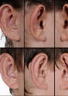
Surgical repair of the earlobe is an increasingly sought-after procedure for patients of all ages. The most common reason for those wanting definitive surgical repair of their earlobe deformity is due to a ‘split’ or elongated earlobe piercing. The resultant cleft or ‘split’ earlobe may be classified as (a) complete or (b) incomplete, the latter referring to continuity of the inferior border of the earlobe remaining intact [1].
There are many causative factors that can lead to enlargement of the initial earlobe piercing which include (a) acute trauma, (b) gauging (the act of placing larger and larger gauge earrings over time), (c) multiple earlobe piercings and (d) persistent wearing of heavy earrings. The ‘split’ earlobe deformity may be exacerbated by the natural effects of facial ageing which will lead to ptosis of the earlobe with increased length and skin creasing.
Although a small structure without specific biological function, the aesthetic appearance of the earlobe is significant. Anatomically, the ear is derived from the second branchial arch and receives its sensory innervation from the greater auricular nerve. Blood supply to the earlobe is supplied by anastomotic connections between the superficial temporal artery (STA) and the posterior auricular artery (PAA), specifically the inferior anterior auricular artery from the STA and the anti-tragal perforator from the PAA [2].
Fortunately, surgical correction of the ‘split’ earlobe is a relatively quick procedure, with minimal complications and one which consistently meets patient expectations. The ideal surgical repair is free from inferior border irregularity / notching, vertical suture line grooving and obvious scarring. Within the literature, many surgical techniques have been described to repair the split earlobe. Some techniques aim to preserve part of the primary piercing, however, in practice, and as is my own preference, most surgical repairs will excise the entire clefted tissue. Grohmann and colleagues have given an excellent systematic literature overview on the various surgical techniques for pierced earlobe repair [3].
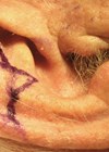
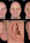
Following a comprehensive patient assessment, the exact surgical technique which I use depends on the size and shape of the split earlobe deformity. The two procedures that I shall describe do not preserve the original earring orifice. The procedure is performed under local anaesthetic (2% lidocaine with a vasoconstrictor) and generally takes under 30 minutes to complete each earlobe. Usually both earlobes are done at the same visit. Next, the edges of the cleft deformity are outlined with a surgical marker. Throughout the procedure, irrespective of repair technique planned, a sterile flat plastic needle container is used to facilitate earlobe position. My preference is to use a size 11 scalpel blade and 6/0 nylon for skin closure with either simple or mattress sutures on both anterior and posterior sides of the earlobe.
- Earlobe repair with incomplete split – the edges around the incomplete cleft can either be ‘punch’ excised (if small <4mm) or excised with the scalpel blade (if >4mm). The refreshed wound edges are sutured together either transversely or vertically – the direction of wound closure determined to minimise inferior border bulging. It is important to note that maintaining the intact natural inferior border of the lobule will aid in minimising split earlobe recurrence.
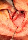
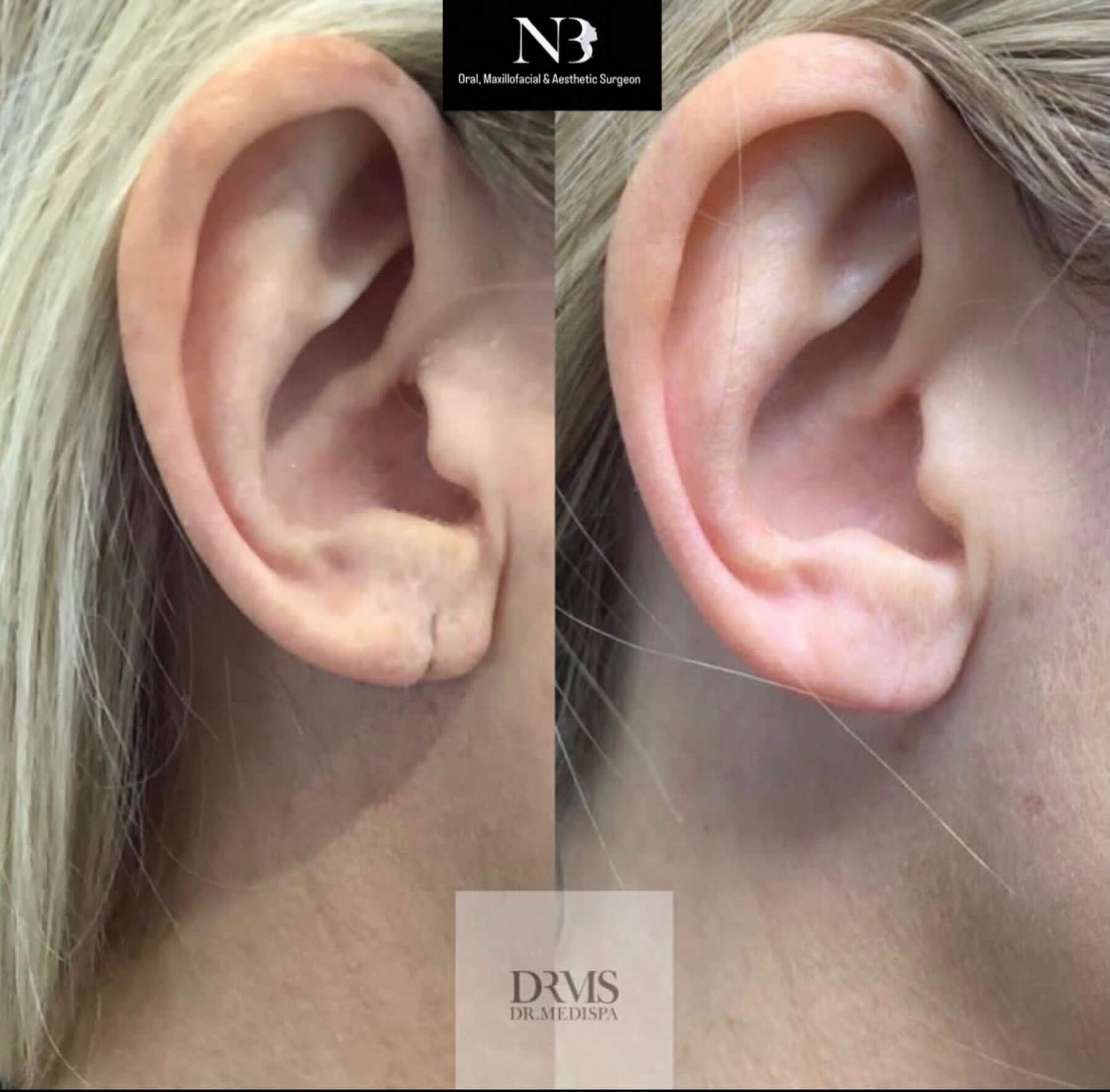
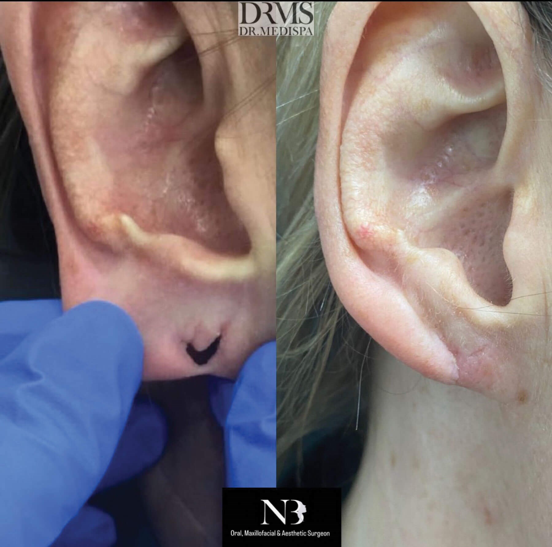
- Earlobe repair with complete split – the inferior border of the earlobe has been completely violated with or without either ptosis or retraction of the split ends. As above, the entire skin of the cleft is marked and excised. If the earlobe is wide and long, I use a simple wedge excision which reduces both dimensions as well as eliminating any deep skin creases present in the ‘ageing’ earlobe. I always aim to incorporate an L-plasty at the inferior end of the earlobe to minimise contour irregularity and thereby enhance the aesthetic result.
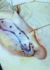
- Earlobe repair with large gauge – this deformity requires reconstruction of both the sizeable hole and ptosis of the earlobe with more complex rotation of the tissue. I reconstruct this type of deformity with a lateral based long pedicle-rotation flap (taking care not to thin the pedicle to less than 2mm thick to ensure good vascularity) which allows for closure of the defect and reduction of the overstretched ‘ptotic’ earlobe tissues.
For those patients whose earlobes are ‘deflated’ in appearance, this may be addressed by injecting a small amount of autogenous fat or dermal filler which markedly rejuvenates the aesthetic appearance.
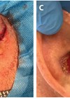
Following surgical earlobe repair, patients are seen for review at six to seven days postoperatively and sutures are removed. There is usually a fine scar when all has healed. Complications (scar retraction, contour irregularities, split lobe recurrence) are minimal when care is taken to choose the best option for split earlobe repair. The procedure generates a high level of patient satisfaction where expectations are managed and patients have been appropriately selected.
References
1. Sadasivan K, Kochunarayanan A. A revised classification and treatment algorithm for acquired split earlobe, with a description of the composite technique and its outcome. Cureus 2020;12(9):e10422.
2. Zilinsky I, Cotofana S, Hammer N, et al. The arterial blood supply of the helical rim and the earlobe-based advancement flap (ELBAF): a new strategy for reconstructions of helical rim defects. JPRAS 2015;68(1):58-62.
3. Grohmann M, Weiland T, Tuca A-C, Wimbauer JM. Earlobe correction of the pierced ear: a systematic review of the literature and principles for surgical reconstruction. Facial Plastic Surgery & Aesthetic Medicine 2022. Epub ahead of print.
Declaration of competing interests: None declared.
COMMENTS ARE WELCOME