We describe our approach to straightforward reconstruction of the peripheral auricle using the established star-pattern design.
Skin malignancies developing on the auricle continue to be regarded as high-risk lesions due to their propensity to recur and for lymphatic spread. Greater peripheral and deep excision margins are therefore required to ensure high rates of complete removal [1,2].
These can produce auricular defects that interfere with the use of spectacles, hearing aids, face masks and other medical devices. Poor cosmesis can result from the altered appearance and asymmetry with the contralateral auricle.
Wedge excision with primary closure is suitable for defects smaller than 15mm in size in the upper and middle third of the auricle. Closure of larger lesions using this approach increases the risk of wound dehiscence and produces distortions in the auricle with anterior bowing. The auricle height is also reduced, although this change is usually subtle. In our experience, wedge excision is rarely suitable when reconstructing the auricle, as lesion sizes and their necessary margins produce considerable defects. Techniques in reconstructing large defects centre on postauricular grafting or a variety of forms of advancement involving skin and cartilage.
We have found the star-pattern advancement reliable in preserving skin, auricular cartilage and form. It is relatively quick to design and perform compared to other types of advancement reconstruction described [3-6]. Scapha tissues are preserved and the shorter operative time makes the reconstruction more tolerable for the patient and suitable to being performed under local anaesthetic.
“We have found the star-pattern advancement reliable in preserving skin, auricular cartilage and form. It is relatively quick to design and perform compared to other types of advancement reconstruction described”
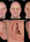
The star-pattern includes three limbs spanning the length of the defect resulting from the initial wide local excision. The length of each limb is no more than 50% of the defect’s length, allowing excellent edge approximation, particularly at the helical rim (Figure 1).
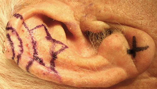
Figure 1: Auricular lesion with excision margin and three-limbed star-pattern design.
The skin is cleaned with 7.5% providone-iodine and skin infiltrated with three to four vials of Lignospan special (lidocaine hydrochloride 2% with 1:80,000 adrenaline). A full pinna block is rarely required. An aperture drape is suitable, as tissue is not being mobilised into the wound from a separate region. A number 15 scalpel blade is used to remove the lesion and margins, with a 3-0 silk marking suture placed at the 12 o’clock position to allow histopathological orientation. A fresh scalpel blade is used to excise the star-shaped pattern from the auricle to avoid cartilage spicules forming that interfere with wound closure.
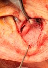
Incisions are made perpendicular to the skin and remove the skin from both the lateral and medial surfaces of the auricle, along with the cartilage to avoid bowing on wound closure (Figure 2).
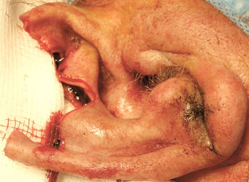
Figure 2: Cartilage and skin from the contralateral surface are removed, facilitating closure and avoiding auricular bowing.
The central limb of the star-pattern is closed initially using 4-0 vicryl rapide sutures, followed by the outer limbs (Figure 3).
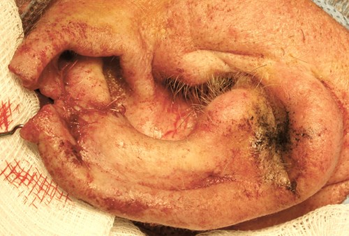
Figure 3: Closure of the central and outer limbs provide good approximation and tension-free closure.
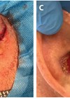
The helical rim is closed with a vertical mattress suture to provide better wound edge eversion, avoiding notching between the advanced superior and inferior limbs of the helical rim (Figure 4).
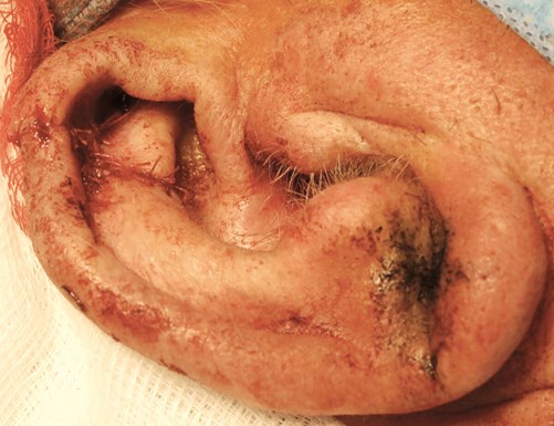
Figure 4: Closure of the helical rim and medial auricular surface completes the reconstruction.
Bactroban ointment is applied to the wound, which is then covered with a Mepore dressing. Formal head bandaging is avoided because of the shearing effect it can apply to the wound. The dressing is removed after five days and the wound reviewed by the operating surgeon seven days post-surgery, with further review one month later.
Whilst a defect size of 25mm has been described as the maximum limit for star-pattern reconstruction, in our experience it can be successfully used to reconstruct defects of any size [7]. The consequence of reconstructing these larger defects is pseudo-microtia but only in cases of extensive defects is this noticeable. In our experience, defects of the inferior third, including the lobule, are suitable for star-pattern reconstruction also. Preservation of even a small part of this subunit permits aesthetically convincing reconstruction.
TAKE HOME MESSAGE
-
The reconstruction has a shorter operative time and is tolerated well by patients under local anaesthetic.
-
The length of each limb is no more than 50% of the defect’s length.
-
Skin is removed from both surfaces of the auricle, avoiding bowing on wound closure.
-
Inferior third defects, including the lobule, are suitable for star-pattern reconstruction also. Preservation of even a small part of this subunit permits aesthetically convincing reconstruction.
References
1. Newlands C, Currie R, Memon A, et al. Non-melanoma skin cancer: United Kingdom National Multidisciplinary Guidelines. J Laryngol Otol 2016;130(S2):S125-32.
2. Ahmed OA, Kelly C. Head and neck melanoma (excluding ocular melanoma): United Kingdom National Multidisciplinary Guidelines. J Laryngol Otol 2016;130(S2):S133-S41.
3. Calhoun KH, Slaughter D, Kassir R, et al. Biomechanics of the helical rim advancement flap. Arch Otolaryngol Head Neck Surg 1996;122(10):1119-23.
4. Butler CE. Reconstruction of marginal ear defects with modified chondrocutaneous helical rim advancement flaps. Plast Reconstr Surg 2003;111(6):2009-13.
5. Dresner H, Waselchuk E. Reconstruction of the middle third auricular defects. Oper Tech Otolaryngol 2017;28(2):114-8.
6. Pickrell BB, Hughes CD, Maricevich RS. Partial ear defects. Semin Plast Surg 2017;31(3):134-40.
7. Antia NH, Buch VI. Chondrocutaneous advancement flap for the marginal defect of the ear. Plast Reconstr Surg 1967;39(5):472-7.
Declaration of competing interests: None declared.
COMMENTS ARE WELCOME