This article has been verified for CPD. Click the button below to answer a few
short questions and download a form to be included in your CPD folder.
Human facial symmetry is a key determinant for assessing facial attractiveness, and achieving a balanced, harmonious, facial appearance is an aesthetic goal that is driving the £24.5 billion aesthetic industry in the UK. Facial asymmetry is an individualised characteristic and is commonly observed sub-clinically in the overall population [1,2].
The extent of facial asymmetry can vary widely from mild to severe and can be measured objectively, but in the author’s experience of having treated over 350 facial disproportion cases, they have found that the patient’s perception is unpredictable, subjective and does not necessarily correlate with objective measurements of severity.
Mandibular asymmetry is one of many facial conditions that impact facial symmetry and balance. The aesthetic and functional consequences of severe mandibular asymmetry can affect social and psychological aspects of quality of life [3–5] and is a common reason why patients pursue treatment. The prevalence of mandibular asymmetry ranges from 17.43–72.95% overall in a systematic review of published samples, and skeletal Class III malocclusion showed the greatest prevalence of mandibular asymmetry (22.93–78%) [6].
Understanding the aetiology of mandibular asymmetry is critical with regards to treatment planning, management and long-term stability. In research settings, the exact criteria to distinguish between pathological and normal asymmetry remains a matter of debate – this is due to a lack of consensus in standardised measurement techniques to address the 3D nature and multiple classification systems in use. However, clinically significant facial asymmetry with associated morphologic, aesthetic, and stomatognathic problems warrant investigation of the underlying aetiology, and comprehensive clinical examination in conjunction with imaging studies for diagnosis, localisation of asymmetry and treatment planning. In the NHS clinical setting it is the functional concerns, whereas in the private sector and aesthetic industry, the aesthetic concerns of mandibular asymmetry that are the primary driving factors for treatment.
Criteria for surgical treatment
Currently, the ‘cut-off’ for state-funded (i.e. NHS) surgical treatment is only possible in individuals assessed with the ‘Index of orthodontic treatment need’ (IOTN) [7] and / or the ‘Index of orthognathic functional treatment need’ (IOFTN) [8] severity scores four and five (i.e. facial asymmetry associated with occlusal disturbance). This leaves many patients with facial asymmetry but no occlusal disturbance turning to the private sector or aesthetic industry (which remains unregulated) for a wide variety of treatments, some of which lack appropriate scientific research evidence support and scrutiny.
Diagnosis of mandibular asymmetry
A full comprehensive assessment of structural, functional and, in some cases, psychological status of the patient by means of thorough history (i.e. presenting concerns, expectations, driving factors, previous facial trauma, relevant medical and psychological history), systematic clinical examination (i.e. objective assessment of extent of asymmetry and exact anatomical structures involved) and appropriate investigations (e.g. study models, occlusal registration or occlusal splints, face-bow transfer, orthopantomogram-OPG) are the essential, indispensable basic requirements for accurate diagnosis of asymmetries. Additional focused investigations (SPECT-CT-scan, electromyogram, CT-scan or MRI-scan) may be needed to establish if asymmetry is progressing or arrested and to help establish underlying aetiology.
Traditionally, skeletal diagnosis of mandibular asymmetry is established, in the sagittal plane, mainly by the location of central points of the mandible such as Pogonion, Gnathion, and Menton. The distance from these central landmarks to the facial ‘mid sagittal plane’ (MSP) is calculated to quantify and classify mandibular skeletal asymmetry as mild (<2 mm), moderate (2–4 mm), or severe (>4mm) [9–11], using cone-beam computed tomography, orthopantomagram, or posterior anterior cephalogram [12].Computed tomography scans allow more accurate assessment of mandibular asymmetry, specifically to establish whether it is roll-dominant (vertical discrepancy), yaw-dominant (horizontal discrepancy) or translation-dominant, which helps to improve treatment planning [13].
Measurement of vertical component of asymmetry
In 1988, Habets, et al. [14] introduced the asymmetry index using orthopantomograms to analyse vertical asymmetries in the mandible, in cases of ramus and / or condylar height asymmetries, which contribute to horizontal / sagittal asymmetries observed. Index values over 6% vertical difference between the right and left sides were considered to have vertical asymmetry, as values less than this are likely due to technical measuring errors.
Various aetiological factors can cause facial asymmetries, such as age, gender, skeletal growth pattern, dental occlusal changes, muscular activities, congenital, developmental and pathological factors. Furthermore, genetic factors can influence the development of facial asymmetries, such as PITX2 and ACTIN3 gene mutations [15]. Personalised treatment planning is the assimilation of numerous factors – patient, medical, aetiological, pathological, structural, functional, occlusal – and objective assessment of the extent of actual asymmetry. Together, these elements inform and aid the clinician in achieving the optimal functional and aesthetic outcome with minimal invasive intervention or side-effects / risks to each individual patient. In the context of jaw surgery, personalised treatment planning is much more than just 3D virtual surgical planning and it is about individualised ‘holistic’ approaches.
Below, the surgeon presents two severe mandibular asymmetry cases with different underlying aetiology and the importance of thorough evaluation and personalised treatment to achieve optimal surgical outcome and meaningful, natural transformation for the patient.

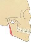
Figure 1: Mid-sagittal plane: Line through nasion, anterior nasal spine and menton. Menton: Most inferior part of the bony chin in the median plane. Gonion: Most posteroinferior point at the angle of mandible. Co: Most superior point of condylar head. Ramus height: Co to gonion.

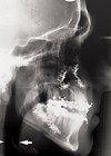
Figure 2a: Case 1 Orthopantomagram: Yaw dominant mandibular asymmetry with chin midline deviation to right.

Figure 2b: Case 2 Orthopantomagram: Roll dominant mandibular asymmetry with no chin midline deviation.
Right ramus height (RRH). Left ramus height (LRH).
Positive asymmetry index (AI) values indicate that the right mandibular ramus is longer; a negative AI indicate an elongated left side. The AI [14] to evaluate the severity of asymmetry between heights of both sides of the ramus of the mandible:
AI, % = RRH – LRH / RRH + LRH × 100%

Figure 3: Pre-op / Post-op.
Case 1
A 20-year-old female presented with functional difficulties (teeth not meeting on the left and only able to chew on a few teeth meeting on the right), temporomandipular joint (TMJ) symptoms, perceived chin prominence and deviation to the right, causing self-esteem and confidence issues.
The potential error in this case would be to treat only the chin (i.e. genioplasty) by correcting the chin asymmetry, as this would not address the underlying pathology (i.e. mandibular asymmetry) and associated dental malocclusion and occlusal cant. Even worse would be to treat the crooked smile and chin asymmetry with injectable fillers as this would not address the underlying skeletal asymmetry.
- Diagnosis: Skeletal base III growth discrepancy, due to maxillary hypoplasia complicated by left hemi-mandibular elongation causing mandibular asymmetry to the right. Yaw dominant mandibular asymmetry (see Figure 1, 2a and 3).
- Aetiology: Developmental growth discrepancy of the mandible.
- Oral assessment: Oral health good. Class 3 malocclusion and lateral open bite left side.
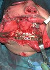
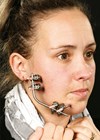
- Treatment: Combined treatment involving pre-surgical orthodontic treatment followed by surgery (bimaxillary osteotomy – Le Fort 1 advancement and asymmetric mandibular rotation to correct asymmetry) and post-surgical orthodontic treatment (see Figure 1, 2a, 3).
Case 2
A 53-year-old female presented with progressive wasting of the left side of her face, crooked smile, perceived facial asymmetry and premature aged appearance of the left side of her face, causing significant self-esteem and confidence issues since her early 20s. Based on IOFTN criteria alone, this patient would not be eligible for state-funded (i.e. NHS) treatment as she did not have occlusal disturbance.

Figure 4: Pre-op / Post-op.
Even though, this lady had similar visible severity of jaw asymmetry to Case 1, jaw surgery treatment would be inappropriate for her as her oral health condition would not withstand the pre-surgical orthodontic (i.e. fixed brace) treatment needed and jaw surgery would not address the volume loss on the left side of face, due to soft tissue atrophy.
- Diagnosis: Parry-Romberg Syndrome (i.e. Left hemifacial atrophy) affecting left side of mandible and soft tissues including muscle and fat. The left ramus height is 5mm shorter compared to the normal right side but no disocclusion due to dental compensation by over-eruption of teeth on left side. Roll-dominant asymmetry (see Figure 2b).
- Aetiology: Unknown – secondary to trauma / hereditary / infection / autoimmune causes postulated.
- Oral assessment: Oral health poor, with chronic periodontal disease. No dental malocclusion.
- Treatment: Liposuction from abdomen and left micro-fat grafting (i.e. facial liposculpture) (see Figure 2b, 4).
Summary
The prevalence of mandibular asymmetry contributing to facial asymmetry is high and can be mild to severe with a wide variety of underlying aetio-pathological causes. Current criteria for state-funded treatment provision does not capture all moderate and severe asymmetry cases, resulting in many patients with mandibular asymmetry seeking treatment in the private sector or the currently unregulated aesthetic industry.
The management of mandibular and facial asymmetry is a highly subspecialised complex area and if misdiagnosed, patients may be subjected to unnecessary extended treatment time or compromised to poor functional and aesthetic outcomes with their consequences, which may be irreversible. The importance of a personalised treatment plan is highlighted to avoid adverse outcomes.
To ensure this group of patients are not subjected to poor safety standards in the currently unregulated aesthetic industry, it is imperative to:
- Improve knowledge and awareness of the public
- Improve sensitivity of criteria used in state-funded service provision
- Develop a fair and comprehensive regulation of aesthetic industry, which will be challenging due to the variety and numerous groups of stakeholders involved, each wishing to protect and preserve their domain of practice.
References
1. Ramirez-Yañez GO, Stewart A, Franken E, Campos K. Prevalence of mandibular asymmetries in growing patients. Eur J Orthod 2011;33:236–42.
2. Melnik AK. A cephalometric study of mandibular asymmetry in a longitudinally followed sample of growing children. Am J Orthod Dentofacial Orthop 1992;101:355–66.
3. McCrea S, Troy M. Prevalence and severity of mandibular asymmetry in non-syndromic, non-pathological Caucasian adult. Annal of Maxillofacial Surg 2018;8(2):254–8.
4. Ryan FS, Barnard M, Cunningham SJ. Impact of dentofacial deformity and motivation for treatment: a qualitative study. Am J Orthod Dentofac Orthop 2012;141:734–42.
5. Soh CL, Narayanan V. Quality of life assessment in patients with dentofacial deformity undergoing orthognathic surgery- a systematic review. Int J Oral Maxillofac Surg 2013;42:974–80.
6. Evangelista K, Teodoro AB, Bianchi J, et al. Prevalence of mandibular asymmetry in different skeletal sagittal patterns: A systematic review. Angle Orthod 2022;92:118–26.
7. Brook PH, Shaw WC. The development of an index of orthodontic treatment priority. Eur J Orthod 1989;11:309–20.
8. Ireland AJ, Cunningham SJ, Petrie A, et al. An index of orthognathic functional treatment need (IOFTN). J Orthod 2014;41:77–83.
9. Thiesen G, Gribel BF, Kim KB, et al. Prevalence and associated factors of mandibular asymmetry in an adult population. J Craniofac Surg 2017;28:e199–203.
10. Masuoka N, Muramatsu A, Ariji Y, et al. Discriminative thresholds of cephalometric indexes in the subjective evaluation of facial asymmetry. Am J Orthod Dentofac Orthop 2007;131:609–13.
11. Damstra J, Fourie Z, De Wit M, et al. A three-dimensional comparison of a morphometric and conventional cephalometric midsagittal planes for craniofacial asymmetry. Clin Oral Investig 2012;16:285–94.
12. Yousefi F, Rafiei E, Mahdian M, et al. Comparison efficiency of posteroanterior cephalometry and cone-beam computed tomography in detecting craniofacial asymmetry: a systematic review. Contemp Clin Dent 2019;10:358–71.
13. Kim HJ, Noh HK, Park HS. Mandibular asymmetry types and differences in dental compensations of Class III patients analyzed with cone-beam computed tomography. Angle Orthodontist 2023;(9):695–705.
14. Habets LL, Bezuur JN, van Ooij CP, Hansson TL. The orthopantomogram, an aid in diagnosis of temporomandibular joint problems. I. The factor of vertical magnification. J Oral Rehabil 1987;14:475‑80.
15. Babczyńska A, Kawala B, Sarul M. Genetic factors that affect asymmetric mandibular growth—A systematic review. Symmetry 2022;14:490.
Acknowledgements
Pamela Ellis (Consultant Orthodontist and Associate Postgraduate Dental Dean for Specialty Training) for peri-operation orthodontic treatment for Case 1; Sian Campbell and Heidi Silk (University Hospitals Dorset NHS Foundation Trust Prosthetic Technicians) for the 3D-VSP images.
Declaration of competing interests: None declared.
COMMENTS ARE WELCOME


