As the incidence of cutaneous malignancy increases further due to both the ageing population and greater disease awareness, the demand for surgical resection within the high-risk regions of the face, including the pinna, is rising [1,2].
Approximately 50% of all cutaneous malignancies of the pinna develop on the helix subunit, including 62% of squamous cell carcinomas [3,4]. The three-dimensional shape of the pinna as well as its complex contouring, significant functional and aesthetic roles pose reconstructive challenges following resection [5]. Whilst offering satisfactory oncological clearance, wedge resections are associated with cupping, webbing from scar formation and microtia [6].
The helical advancement flap is an established method of reconstruction for the entire helical subunit and can be extended to the scapha due to its composite chondrocutaneous structure. The flap is axial in nature, receiving reliable blood supplies from the anterior auricular artery to the helix and superficial temporal artery to the lobule. We perform a variant of the flap originally described by Antia & Buch (1967), which is quicker to complete and can be used to manage the largest defects, including those emanating from the upper portion of the superior third, through creation of a superior limb [7].
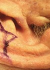
The lesion’s edge and excision margins are marked prior to infiltration with Lignospan® Special (lidocaine hydrochloride 2% with 1:80,000 adrenaline), both to increase the volume of local anaesthetic that can be safely administered and to reduce bleeding from the vasoconstrictive effect. The infiltrated area of the pinna is then digitally massaged to promote infiltration into the soft tissues and provide a degree of hydro-dissection of the tissue plains. The skin is cleaned with povidone-iodine 7.5% and covered with a simple aperture drape when dried as tissue distant to the pinna is uninvolved. Once the lesion has been excised and appropriately orientated, the wound edges on the posterior pinna surface are connected by an acute triangle design and the skin and underlying cartilage of this is excised to allow an O-T closure during advancement of the helix (Figure 1).

Figure 1.

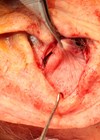
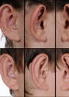
Figure 2.

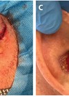
Figure 3.

Figure 4.

Figure 5.
The helical advancement flap is almost always inferiorly-based. An incision is made at the junction of the helix and scapha using a No.15 scalpel blade. The incision follows the curvature of the pinna inferiorly towards the superior aspect of the pinna lobule (Figure 2). The incision can be placed more medially as determined by the size of the primary defect. Whilst the incision divides the anterior pinna skin and cartilage, it is essential that the underlying skin of the posterior pinna is preserved throughout the operation to maintain the blood supply to the flap from the branches of the anterior auricular artery. Division of the posterior pinna skin increases the risk of cartilage necrosis with subsequent loss of strength and poor cosmesis.
A single Burow’s triangle is created by lobule skin excision (Figure 2). No fat removal is necessary as its own compressibility and the skin laxity over this subunit allow easy advancement superiorly. The anterior skin overlying the cartilage is separated off by sharp dissection using a fresh No.15 blade (Figure 3). The skin remains elevated using skin hooks whilst a 3mm sliver of scaphal cartilage is also removed by sharp dissection (Figure 4). It is these excisions of skin and cartilage that provide sufficient space for advancement and tension-free closure without anterior cupping or loss of pinna height leading to asymmetry with the contralateral ear.
The wounds are closed with interrupted sutures using 4-0 vicryl rapide material (Figure 5). The incisions are concealed well within the curvatures of the lateral pinna and its posterior surface. The wounds are covered with Bactroban ointment (mupirocin 2%) and a Jelonet dressing (paraffin-soaked gauze) to maintain moist, antiseptic surfaces. The pinna is covered with cotton gauze swabs anterioly and secured with a head bandage, principally to protect the operative site rather than to provide compression. This is removed the following morning after surgery. This technique of reconstruction can be applied following keloid excision as well following trauma and burns to the area if soft tissue is lost.
References
1. Venables ZC, Nijsten T, Wong KF, et al. Epidemiology of basal and cutaneous squamous cell carcinoma in the UK 2013-15: a cohort study. Br J Dermatol 2019;181(3):74–482.
2. Goon PK, Greenberg DC, Igali L, Levell NJ. Predicted cases of UK skin squamous cell carcinoma and basal cell carcinoma in 2020 and 2025: horizon planning for National Health Service dermatology and dermatopathology. Br J Dermatol 2017;176(5):1351–3.
3. Sanniec K, Harirah M, Thornton JF. Ear reconstruction after Mohs cancer excision: lessons learned for 327 consecutive cases. Plast Reconstr Surg 2019;144(3):719–29.
4. Freelander E, Chung FF. Squamous cell carcinoma of the pinna. Br J Plast Surg 1983;36(2):171–5.
5. Noel W, Leyder P, Quilichini J. Modified Antia-Buch flap for the reconstruction of helical rim defects. J Plast Reconstr Aesthet Surg 2014;67(12):1659–62.
6. Majumdar A, Townend J. Helix rim advancement for reconstruction of marginal defects of the pinna. Br J Oral Maxillofac Surg 2000;38(1):3–7.
7. Antia NH, Buch VI. Chondrocutaneous advancement flap for the marginal defect of the ear. Plast Reconstr Surg 1967;39(5):472–7.