To non-radiologists ultrasound may always look like a snowstorm . . . so unless you are the one holding the scanner and moving it over the patient with some idea of their history, it is difficult to know what you are looking at.
Within the last five years, point-of-care ultrasonography (POCUS) has become a realistic and affordable option in healthcare. For £5k or so, technological advances in miniaturisation have shrunk these units to be not much bigger than a smartphone, and to transmit images to an ordinary phone or tablet via WiFi.
As a plastic surgeon in full-time aesthetic practice, there are a large number of potential applications for POCUS. From the outset it is very important to advise patients that you are not a radiologist or radiographer and that the scan is being undertaken in order to give a general idea of the underlying anatomy, not as a substitute for formal radiological examination, which of course represents the gold standard. It goes without saying that patients with unexpected or suspicious findings should always be referred. We are merely using this excellent technology to inform decisions that we would otherwise have made in the operating room or the clinic with far less information to hand about what we cannot see under the skin. Point-of-care ultrasonography enhances clinical decision-making.


Figures 1–3: Before and after scans of post abdominoplasty seroma.
Applications in clinical practice
Preoperatively, subcutaneous fat deposits can be measured in liposuction, lipoedema or Brazilian butt lift (BBL) cases. It is now possible to measure changes with a high degree of accuracy rather than by photographing or surface scanning the external visual change.
In breast augmentation consultations upper pole soft tissue coverage can be assessed and measured more precisely than with a calliper, which can sometimes inadvertently include pectoralis major muscle in the pinch measurement, a vital metric for choosing whether to place the implant in front of or behind the pec major muscle. Implanted breast prostheses can also be assessed for orientation (in the case of suspected rotation or flipping of anatomical / teardrop shaped implants). Breast implant shells can also be scanned to check their integrity in cases of suspected rupture. Of course, the gold standard test for implant shell integrity is MRI however POCUS can raise an index of suspicion and allow onward referral for MRI.
Intracapsular fluid collections can also be seen and aspirated allowing earlier detection of breast implant-associated anaplastic large cell lymphoma (BIA-ALCL).
Intraoperatively, ultrasound has revolutionised the safety of gluteal fat transfer (BBL) by allowing the operating surgeon to see the exact plane where the fat is being placed in real time (Figure 1) – removing guesswork and keeping the cannula within the fat above the gluteus maximum muscle massively reduces the chances of fatal fat embolism as the largest veins which are greatest risk for fat embolism lie within the muscle. This has been proposed as a mandatory use in this procedure.


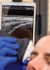
Figures 4–5: Ultrasound in practice by Lauren Mackintosh, Advanced Aesthetics Practitioner at Wood MediSpa.
Postoperatively, fluid collections are very easy to see and drain. Abdominal seromas or collections around breast implants are either there or they’re not! Ultrasound enables you to see the difference between soft tissue swelling and a seroma. It is possible to mark the full extent of a seroma with a skin marker, aspirate it and then rescan the area post aspiration to ensure the space is completely empty.
The needle enhance mode makes it easy to see a hypodermic needle and direct it towards a seroma and POCUS alleviates any concerns about inadvertently entering the peritoneum or bladder during abdominoplasty seroma aspiration.
In terms of facial aesthetics and safety of injectable fillers much has been written on POCUS in this role already. A different handpiece with a higher frequency is necessary for facial imaging as the required depth of penetration is far less than with soft tissue scans of the body and limbs.
Pre-filler treatment vascular mapping using colour Doppler can demonstrate the location and depth of the facial arterial supply and massively reduce the risk of vascular occlusion (VO) particularly in conjunction with blunt cannula techniques in high-risk areas of the face. If VO does occur then the colour Doppler can demonstrate the location of the occlusion and help to guide hyaluronidase injections.
Recently I had a patient return to me several months post facelift and facial fat transfer with a couple of tiny nodules in the areas where her fat had been grafted. I scanned her and saw two tiny fluid filled cysts. These were easy to aspirate under local anaesthetic using a hypodermic needle and completely disappeared. The issue was diagnosed and fixed in a couple of minutes and the patient left happy.
In my opinion this technology is here to stay. It has a multitude of potential applications and provides helpful insight informing clinical decision-making. The only way to become competent at using it is to invest in it and use it regularly. Further information British Medical Ultrasound Society (BMUS). Guidelines for professional diagnostic ultrasound practice in medical aesthetics.
Declaration of competing interests: None Declared.
COMMENTS ARE WELCOME