The upper face, including forehead and temples, is often overlooked in non-surgical cosmetic procedures with dermal fillers. While horizontal forehead or glabella lines may be a common complaint amongst patients, seldom do they attend with concerns of loss of volume or contouring in the upper face.
As profiloplasty and three-dimensional facial analysis become more common, attention is gradually focusing on this region, making it an important area for consideration in any rejuvenation and aesthetic treatment plan.
Despite the potential for harmonious outcomes, many clinicians may feel apprehensive about approaching this area due to its complex anatomy and the risk for serious complications, such as vascular occlusion that could lead to skin necrosis and blindness. This article will explore the intricate anatomy of the upper face focusing on the layered arrangement, vascularisation, discuss safer injection techniques using hyaluronic acid dermal fillers, and emphasise the importance of prioritising patient safety in cosmetic procedures.

Figure 1: Forehead and temple boundaries.
Boundaries
When treating any facial region, it is crucial to recognise its boundaries and topographical landmarks as these are key references for injections (Figure 1).
The forehead is the area defined:
- Superiorly by the hairline, which is more of a clinical or aesthetic boundary than an anatomical one.
- Inferiorly by the supraorbital rim, with the aesthetic boundary being the eyebrows.
- Laterally on both sides is the temporal crest, this can quite often be visible and palpable.
The temporal area is defined:
- Superiorly by the temporal crest.
- Inferiorly by the superior border of the zygoma.
- Anteriorly by the lateral external border of the orbital rim.
- Posteriorly by the temporal hairline, considered a clinical not an anatomical boundary.
Ageing and ideal shape
Ageing of the upper face is a multifaceted process that involves a complex interplay of all facial layers including fat compartments, bone, retaining ligaments, musculature, and skin. This process is not consistent across individuals, as factors such as genetics, lifestyle, and environmental influences can all impact the rate and severity of ageing signs [1].
One of the most noticeable changes is the loss of volume in the central and lateral aspects of the forehead accompanied by temporal hollowing. Additionally, alterations in the positioning of the eyebrows can occur leading to a depressed appearance.
Volume deflation of the temporal fossa can lead to a skeletonised appearance with an emaciated look to the upper face and accentuated bone prominences of the lateral orbital rim and zygomatic arch. Other visible signs are the outline of the temporal crest and vasculature. However, this skeletonisation is not always age-related and can also be present in young patients.
Restoring volume and shape can address some of these changes, but one should not overlook important factors such as gender and ethnic background. For example, in Asia, a very rounded forehead and temples are considered more attractive while for Caucasians a flat or discreet convexity is typically preferred. Male patients have a more pronounced supraorbital bossing therefore presenting with a flat area before the convex curvature of the upper forehead begins. In the female, the supraorbital bossing is usually less or quite often non-existent, leading to a more continuous mild curvature [2,3].
In general, there is no ideal shape nevertheless a youthful and balanced upper facial contouring shows a gentle convexity of the forehead with a smooth transition to the temples, extending to the lateral midface.
Layered arrangement of the forehead and temples
A recent study re-evaluated the anatomy of the forehead using cadaveric dissections and ultrasound imaging of live subjects, revealing a novel concept by identifying additional layers (suprafrontalis fascia, retrofrontalis fat and subfrontalis fascia) to the already recognised layered arrangement. Additionally, reinforcing the deep fat compartments or supraperiosteal plane as the preferable location for dermal filler injections to address loss of volume and contour, as nerves and arteries travel deep to the frontalis muscle but superficial to the subfrontalis fascia [4]. The temple, also referred to as the temporal fossa, has a more intricate anatomical arrangement compared to other facial areas.
Current literature shows the presence of 13 layers [5], all already described by previous cadaveric studies [6] but with a somewhat different nomenclature. The superficial temporal fascia (STF) encases the superficial temporal artery within its laminae (layers 3 and 4) and it represents the superficial musculoaponeurotic system (SMAS) in this region.

The deep temporal fascia (DTF) separates into a superficial (layer 8) and a deep layer (layer 10) before their connection to the zygomatic arch, enveloping layer 9 (superficial temporal fat pad).The deep temporal fat pad that lies under the DTF is also known as temporal extension of the buccal fat pad of Bichat. If injections are too low in the temporal fossa, the product can spread to the midface via this fatty connection.
Appreciation of this complex layered arrangement is essential when selecting the injection technique; more superficial injections with a less cohesive product are indicated for light to mild correction, and deeper delivery of the material such as in supraperiosteal for correction of moderate to severe depression of the temple require a higher G prime product. Deep injections also require a greater amount of product to fill the depression and ‘lift’ the overlying layers. A combination of techniques can provide a better clinical outcome in selected cases [7].
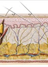
A Brazilian study from 2023 questioned the safety of interfascial injections after a high incidence of incorrect placement (89%) when these were performed with no image guidance in fresh frozen specimens, recommending the use of ultrasound imaging when selecting this technique [8]. A detailed ultrasound image illustrates the intricacy of this layered arrangement (Figure 2).

Figure 2: Ultrasound image of layered arrangement of forehead and temples.
Credit: Jair Mauricio Cerón Bohórquez, MD, Medical Contour, Hamburg, Germany.
Vascular supply
The plethora of anastomosis or connections between the internal carotid artery (ICA) and external carotid artery (ECA) is the main reason this is a treacherous and risky area for adverse events if accidental intravascular injection occurs.
A. Supratrochlear (STA) and supraorbital (SOA) arteries:
The STA and SOA are both terminal branches of the ophthalmic artery, which itself is a branch of the ICA, providing the primary vascular supply to the forehead.
The STA emerges at the lower medial margin of the orbital rim, piercing the corrugator muscle, with superficial and deep branches supplying the corresponding aspects of the frontalis muscle. The SOA exits from the supraorbital foramen or notch at the lower margin of the orbital rim, runs deep to the frontalis, then pierces it to run superficially. One study demonstrated variations in the distribution pattern of these vessels in the forehead, with the deep branch of the STA occasionally not present [9].
After investigating 50 patients using ultrasound imaging, Cotofana et al. confirmed that these arteries changed plane from deep to superficial at a mean distance of 13-14mm from the superior orbital rim. Notably, they demonstrated that both arteries course deep to the orbital part of the orbicularis oculi muscle after emerging from their respective foramen / notch and not in direct contact with the periosteum. They provided clinically relevant information for clinicians to perform safer frontal procedures by suggesting that injections for the upper forehead should target the deep supraperiosteal layer as vessels are located in a more superficial plane here. Furthermore, they indicate that soft tissue fillers can also be administered into the supraperiosteal plane in the lower forehead with a certain degree of safety [10].
B. Central and paracentral arteries:
Special attention should be given to these vessels as they supply the inferior and middle third of the central forehead, a common site for vascular complications due to the rich anastomosis with the angular and supratrochlear arteries.Although not always present or identifiable, they are recurrently found within the superficial fat layer, therefore injections in the glabella should be performed by using intradermal serial punctures [11,12].
C. Superficial temporal artery:
This is a terminal branch of the ECA, located within the pliable superficial temporal fascia and it has an important direct connection by its frontal branch to the supratrochlear and supraorbital artery of the forehead. Injections in the temple region should avoid entering this fascia.
D. Deep temporal arteries (DTA) and middle temporal arteries (MTA):
These are important vessels to consider when performing deep temporal injections. The anterior and posterior DTAs are branches of the internal maxillary artery. In a cadaver study, the DTAs were identified bilaterally in all specimens, within the temporalis muscle and superficial to the periosteum [13]. The anterior branch has a direct connection to the lacrimal artery (branch of the ophthalmic artery) via the zygomatico-temporal artery, posing a risk of adverse events if intravascular injections occur. The posterior DTA connects to the zygomatico-orbital and the anterior DTA. Vascular complications can take place due to the zygomatico-orbital connections with the transverse facial artery, the zygomatico-facial artery, and the superficial temporal artery. The MTA originates from the STA and has connections with the DTAs, therefore it is also vulnerable to iatrogenic intravascular injections and complications. The deep and middle arteries pass within the deep portion of the temporalis muscle, decreasing in diameter as they ascend the fossa [7].

On account of the high variability of location and branching patterns of the DTAs, it is improper to locate a safe zone of injection for deep temporal injections with needles.
Recommended injection techniques for forehead and temples should be based on anatomical evidence found in the literature, respecting danger zones and ensuring the best outcome (Table 2).
Conclusion
While the upper face may present unique challenges in terms of anatomy and potential risks, it is incumbent upon us to prioritise our patients’ safety above all else when considering interventions in this region. Appropriate training and sound knowledge of anatomy will allow practitioners to deliver effective treatments that rejuvenate and beautify while minimising the likelihood of complications.
References
1. Cotofana S, Mian A, Sykes JM, et al. An update on the anatomy of the forehead compartments. Plast Reconstr Surg 2017;139(4):864e–72e. doi:10.1097/PRS.0000000000003174
2. Frank K, Gotkin RH, Pavicic T, et al. Age and gender differences of the frontal bone: a computed tomographic (CT) based study. Aesthet Surg J 2019;39(7):699–710.
3. Dempf R, Eckert AW. Contouring the forehead and rhinoplasty in the feminization of the face in male-to-female transsexuals. J Craniomaxillofac Surg 2010;38(6):416–22.
4. Ingallina F, Frank K, Mardini S, et al. Reevaluation of the layered anatomy of the forehead: introducing the subfrontalis fascia and the retrofrontalis fat compartments. Plast Reconstr Surg 2022;149(3):587–95.
5. Ingallina F, Alfertshofer MG, Schelke L, et al. The fascias of the forehead and temple aligned – an anatomic narrative review. Facial Plast Surg Clin North Am 2022;30(2):215–24.
6. Cotofana S, Gaete A, Hernandez CA, et al. The six different injection techniques for the temple relevant for soft tissue filler augmentation procedures – clinical anatomy and danger zones. J Cosmet Dermatol 2020;19(7):1570–9.
7. Ingallina F. Facial Anatomy and Volumizing Injections 2017 (Superior and Middle Third). Archidemia; 2017.
8. de Lima Faria GE, Nassif AD, Schwartzmann G, et al. Interfascial technique for volumizing the temple with no image guidance: is it safe? Eur J Plast Surg 2023;46:717–23.
9. Cong LY, Phothong W, Lee SH, et al. Topographic analysis of the supratrochlear artery and the supraorbital artery: implication for improving the safety of forehead augmentation. Plast Reconstr Surg 2017;139(3):620e–7e.
10. Cotofana S, Velthuis PJ, Alfertshofer M, et al. The change of plane of the supratrochlear and supraorbital arteries in the forehead-an ultrasound-based investigation. Aesthet Surg J 2021;41(11):NP1589.
11. Scheuer JF 3rd, Sieber DA, Pezeshk RA, et al. Facial danger zones: techniques to maximize safety during soft-tissue filler injections. Plast Reconstr Surg 2017;139(5):1103–8.
12. Koziej M, Polak J, Hołda J, et al. The arteries of the central forehead: implications for facial plastic surgery. Aesthet Surg J 2020;40(10):1043–50.
13. Nikolis A, Enright KM, Troupis T, et al. Topography of the deep temporal arteries and implications for performing safe aesthetic injections. J Cosmet Dermatol 2022;21(2):608–14.
14. Pirayesh A, Bertossi D, Heydenrych I. Aesthetic Facial Anatomy Essentials for Injections. CRC Press; 2020.
Declaration of competing interests: None declared.
COMMENTS ARE WELCOME


