In recent years, there has been a shift in how patients see themselves. The ‘selfie generation’ are constantly exposed to idealised images, which means that many patients are hyper-aware of subtle facial asymmetries and textural irregularities. They no longer look at their faces in static mirrors – they evaluate themselves in motion, under different lighting, and through a highly curated lens.
This shift has created both opportunities and challenges for aesthetic doctors. On one hand, patients are more motivated to seek treatments; on the other, their expectations can be based on unrealistic standards. Our role is to bring them back to reality, to help them understand beauty through a clinical, anatomical lens rather than a digital one. This is where Aura 3D imaging technology can help transform the patient experience in my practice.
First steps and consultation
Aura helps me design bespoke treatment plans tailored to the patient’s anatomy, proportions and goals. By visualising the face in 3D, I can show how volume restoration or contour refinement would appear from multiple angles. This transparency builds trust and turns the consultation experience from ‘doctor telling’ to ‘doctor and patient co-creating’.
Many new patients mistakenly believe that a single treatment will ‘fix’ everything, others misjudge their problem areas; for example, they may think they need nasolabial filler when in fact the issue is midface support loss.

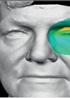
Three-dimensional imaging allows me to objectively demonstrate these misconceptions, helping patients visualise the cause of their concern. While 2D photos flatten the anatomy, missing shadow transitions, contour depth, and proportional relationships, 3D imagery lets the patient view their face from multiple perspectives, shifting confusion into clarity. This level of interactivity turns the consultation into a discovery process where patients are more engaged.
In an era saturated with idealised imagery, this realism is crucial. With Aura, I can ground every discussion in the patient’s actual anatomy. I can show them ideal scenarios and discuss their likely trajectory based on tissue quality, age and skin response. The 3D visuals help bridge that gap, turning aspiration into actionable realism.
This immersive experience directly influences decision-making; patients see potential outcomes in a realistic, data-driven way, fear is replaced by confidence. This is not about ‘selling’ a procedure, it is about empowering patients with insight.
The technology enhances how I communicate treatment limitations; for instance, showing when severe laxity cannot be fully corrected non-surgically. It provides a visual language that patients intuitively understand. Instead of abstract descriptions, I can point to specific volumetric deficits, asymmetries or areas of descent. That honesty, backed by visual evidence, sets the right expectations early.
During and post-treatment
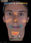
For patients undergoing multi-step or long-term treatment plans, Aura serves as a visual roadmap, and I use scans to track outcomes over time. The 3D overlay feature makes progress visible, showing incremental volume gains after biostimulatory treatments or improvements in symmetry after energy-based remodelling. This objective tracking reassures patients that improvement is happening, even when it is gradual. Patients can see their transformation unfold, strengthening retention, trust and long-term satisfaction.
When viewing their results in 3D versus 2D, patients respond differently; they appreciate nuances they might miss in 2D photography, such as the harmony of their contours, not just isolated changes. Emotionally, it shifts the reaction from “Wow, that line is gone” to “Wow, my whole face looks more balanced.” Importantly, the technology can reduce miscommunication between practitioner and patient. Many misunderstandings stem from unreal expectations. By showing patients both baseline and simulated results, we align on the same visual endpoint. There is no ambiguity; we are both looking at the same face, from the same perspetive.
Conclusion
Ultimately, 3D visualisation is increasingly integral for patient education and for treatment validation and research. Quantifiable change (not just photographic evidence) is becoming the new gold standard. In the future, AI-driven analytics layered onto 3D scans could help predict treatment responses and customise protocols based on the patient phenotype.
The technology is a communication and documentation tool, and a trust-building platform, which can transform patient relationships, treatment planning and results.
Declaration of competing interests: None declared.