Fat necrosis can occur at any site in the body; the majority of cases are in the breast and are idiopathic. Other aetiologies include trauma, fine needle aspiration cytology (FNAC), both cosmetic and therapeutic surgical procedures, autologous lipofilling of the breast and breast irradiation [1].
It can present as a painful mass, which may resolve or remain as an oil cyst; radiological imaging and core biopsy are crucial for an accurate diagnosis [2–4].
Covid-19 infection has been associated with epicardial fat necrosis [5] and it can lead to fat embolism [6] and bone marrow necrosis [7]. The relationship between fat and skin necrosis and the administration of the Covid-19 vaccine, has been recently highlighted in a small number of case reports. Facial fat necrosis [8], toxic epidermal necrolysis [9] and cutaneous thrombosis [10], have all been documented as occurring shortly after immunisation with the Covid-19 vaccine, and are postulated to be vaccine-induced thrombotic events [9]. To our knowledge, to date, there has been no report of fat necrosis of the breast associated with the Covid-19 vaccination.
Case report
A previously healthy, 72-year-old female, with no prior history of breast disease or anticoagulant use, presented with bilateral, exceptionally painful breast lumps, a week following the first dose of the Astra Zeneca Covid-19 vaccine. She had initially noted a local reaction at the injection site, followed by a viral-like illness. Her Covid-19 PCR test was negative, and her c-reactive protein (CRP) was significantly elevated at 170 units (normal <5mg/L). There was no history of breast trauma, surgery or irradiation. Clinically she had multiple bilateral painful breast masses; mammography and MRI were both negative and ultrasound was reported as U2, with bilateral, patchy diffuse increased reflectivity, suggestive of extensive mastitis (Figure 1).

Figure 1: Left breast ultrasound scan reporting inflammatory changes. Chest X-ray and CT revealed mild inflammatory changes and a core biopsy of one of the typical breast lesions demonstrated fibrofatty breast parenchyma with foamy histiocytes, mild oedema and dilated capillaries (U2), in keeping with a diagnosis of fat necrosis (Figure 2).

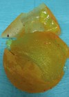
Figure 2: Histology demonstrated changes consistent with the diagnosis of fat necrosis of the breast.
Treatment was conservative and at follow-up three months later, all the masses had reduced in size and were completely pain free. She was discharged with the advice to remain ‘breast aware’.
Discussion
Fat necrosis of the breast is a benign non-suppurative inflammatory process, which results in aseptic saponification of the breast fat and is commonly seen in female patients who have undergone surgical procedures to the breast, including cosmetic interventions [1]. These comprise breast reduction, mastopexy, fat grafting and autologous tissue breast reconstruction. Other causes can include breast trauma, FNAC, anticoagulant therapy, therapeutic breast surgery and breast radiotherapy.
The condition can result in an oil cyst or a breast lesion, which can mimic malignancy, both clinically and radiologically [5]. The patient in this case had no previous history of breast trauma and was not on anticoagulants. All presenting patients should be discussed at the breast multidisciplinary team (MDT) meeting and require a full clinical examination, appropriate imaging and core biopsy to confirm the diagnosis of fat necrosis, which carries an excellent prognosis. Treatment can be conservative, as in this case, or may include the aspiration of oil cysts or even surgical excision if there is no resolution of the pain or breast distortion is present.
In the published literature, there are a small number of case reports of fat necrosis following Covid-19 infection [5–7), and more recently a few reports of skin, fat and soft tissue necrosis have appeared in a variety of medical publications. In 2022, Brau-Javier and colleagues described facial fat necrosis and induration of the facial skin, subsequent to aesthetic facial surgery with autologous fat transfer, followed by Covid-19 vaccination at one-week post-surgery. Their conclusion was that the vaccine had initiated a thrombotic event leading to ischemia of the soft tissues, resulting in fat necrosis, as the cutaneous induration extended over most of the cheek [8].Guarino and colleagues in 2024 successfully treated a case of toxic epidermal necrolysis, which came on five days following the Covid-19 vaccine and postulated that the mRNA vaccine had initiated immune-thrombosis [9,10]. Cutaneous thrombosis following the Covid-19 vaccination has also been described by Gallo-Pineda and colleagues [11], and they have highlighted the risk of rare Covid-19 vaccine-associated thrombotic episodes.
To date there appears to be no documentation in literature, of the potential association of the Covid-19 vaccination with extensive fat necrosis of the breast. We postulate that in this case, the vaccination, one week prior to the clinical symptoms appearing, was the causal factor which affected the probability [12] of there being a widespread thrombotic episode involving the breast adipose tissues. This strong relationship between exposure to the vaccine and the outcome (extensive fat necrosis of the breasts) robustly suggests a greater probability of causation. It is our wish to alert clinicians of different specialties, who are involved in breast surgery, of this probable connection, the final diagnosis of fat necrosis only being made after a full clinical, radiological and pathological examination of the patient.
Conclusion
Although an association of events is not absolute proof of cause, we wish to draw the attention of our colleagues to the potential connection between the Covid-19 vaccine and fat necrosis of the breast and we would welcome any other such cases being brought to our attention.
References
1. Genova R, Garza RF. Breast fat necrosis. StatPearls [Internet]. 2025.
https://pubmed.ncbi.nlm.nih.gov/31194348/
[Last accessed 27 August 2025).
2. Kerridge WD, Kryvenko ON, Thompson A, Shah BA. Fat necrosis of the breast: a pictorial review of the mammographic, ultrasound, CT, and MRI findings with histopathologic correlation. Radiol Res Pract 2015:613139.
3. Dillon MF, Quinn CM, McDermott EW, et al. Diagnostic accuracy of core biopsy for ductal carcinoma in situ and its implications for surgical practice. J Clin Pathol 2006;59(7):740–3.
4. Park HL, Hong J. Vacuum-assisted breast biopsy for breast cancer. Gland Surg 2014;3(2):120–7.
5. Zayour M, Karaki M, Al Ashkar R, Chammas E. Epicardial fat necrosis after Covid-19 infection: a case report. Cureus 2022;14(5):e25154
6. Cinti S, Graciotti L, Giordano A, et al. Covid-19 and fat embolism: a hypothesis to explain the severe clinical outcome in people with obesity. Int J Obes (Lond) 2020;44(8):1800–2.
7. Ghosh S, Gupta SS, Mehta N, Khodaiji S. Covid-19–associated bone marrow necrosis – a case report. Indian J Radiol Imaging 2021;31(03):725–8.
8. Brau‐Javier CN, Caro‐Muniz AP, Canizares O. Facial fat necrosis after autologous fat transfer possibly associated with SARS‐CoV‐2 vaccine. J Cosmet Dermatol 2023;22(5):1477–80.
9. Guarino M, Benedetto M, Blanaru OT, et al. A severe adverse event after COVID-19 vaccination: a case report of toxic epidermal necrolysis syndrome. J Emerg Crit Care Med 2024;8:4.
10. Ramessur R, Saffar N, Czako B, et al. Cutaneous thrombosis associated with skin necrosis following Oxford‐AstraZeneca Covid‐19 vaccination. Clin Exp Dermatol 2021;46(8):1610–12.
11. Gallo‐Pineda G, Navarro‐Navarro I, Viedma‐Martínez M, et al. Skin necrosis after Covid‐19 vaccines. J Dtsch Dermatol Ges 2022;20(12):1654–6.
12. Dhawan R, Shay D. Principles of Causation. StatPearls [Internet]. 2024.
https:/www.ncbi.nlm.nih.gov/books/NBK606119/
[Last accessed 27 August 2025].
Declaration of competing interests: None declared.