The link, both historical and contemporary, between art and anatomy is apparent. A recent British Broadcasting Corporation (BBC) documentary The Beauty of Anatomy showcased the impressive contributions of artists towards the deeper understanding of anatomy and, by default, surgical practice. Such eminent artists and anatomists included the likes of Claudius Galen, Leonardo Da Vinci, Andreas Vesalius, the Hunter brothers and Henry Vandyke Carter (of Gray’s Anatomy fame) [1].
Drawing as a form of an anatomy teaching tool has been adopted in France for many years, being delivered through ‘blackboard and chalk’ drawing sessions. These allowed tutors to demonstrate anatomical planes and structures in a clear and sequential manner, and these were often easier to understand and visualise compared to prosections and cadavers for students [2]. In recent years, the UK’s medical curriculum has transformed and has shifted toward problem-based learning and patient-centre objectives, often at the expense of anatomy. In fact, a national survey has suggested that an important reason behind why many medical students did not intend to pursue a career in surgery was due to a lack of confidence in their anatomical knowledge, which may strongly be attributed to insufficient teaching at the undergraduate level [3].
“Drawing helps students to engage with the complex anatomical planes, thereby allowing them to approach cadaveric dissection with sufficient prior knowledge and to subsequently consolidate it effectively.”
We are not suggesting that the incorporation of drawing into the anatomy curriculum would compensate for the reduction in dictatorial and cadaveric anatomical teaching, but we do believe in its utility as a tool that teachers and students can adopt into their teaching and learning. This article focuses on the teaching of head and neck anatomy through the medium of drawing, as it is a complex region which students often find the most difficult to visualise and understand.
Head and neck anatomy teaching session
We designed a one-day head and neck anatomy teaching session for undergraduate students studying medicine, dentistry or science degrees. Our goal was to improve their knowledge of head and neck anatomy, not only through didactic, factual teaching, but also by equipping them with drawing techniques that would assist them in retaing their knowledge of anatomy more effectively. The session was delivered in the university dissection room by the two authors of this article, both qualified doctors and anatomy demonstrators with a keen interest in medical illustration. The sessions were taught through a combination of ‘white-board’ drawing demonstrations and dictatorial teaching, as well as a conclusive cadaveric demonstration at the end to consolidate their knowledge. Students were provided with pencils and paper to draw throughout the drawing demonstration, and were given a booklet of pre-drawn anatomical structures for labelling as required. Due to strict laws governing dissection rooms, photographs were not allowed to be taken during the session..
Figures 1-5 represent some of the illustrations that were used during the drawing demonstrations and which were included in the work booklet. The topics that were covered included: the facial skeleton and associated sinuses, muscles of facial expression and mastication, the anatomical pathway of the facial and trigeminal nerves, the nasal and oral cavities, the larynx and vocal cords, neck compartments, and basic head and neck embryology. These above topics were selected for their clinical relevance, which included common facial fractures, facial nerve palsy, dental infections, vocal cord palsy, and surgical neck dissections. We were aware of certain topics that could not be covered due to time constraints, such as neuroanatomy, the skull base, orbital anatomy, the auditory system and other more specific topics such as the temporal and infratemporal fossa.
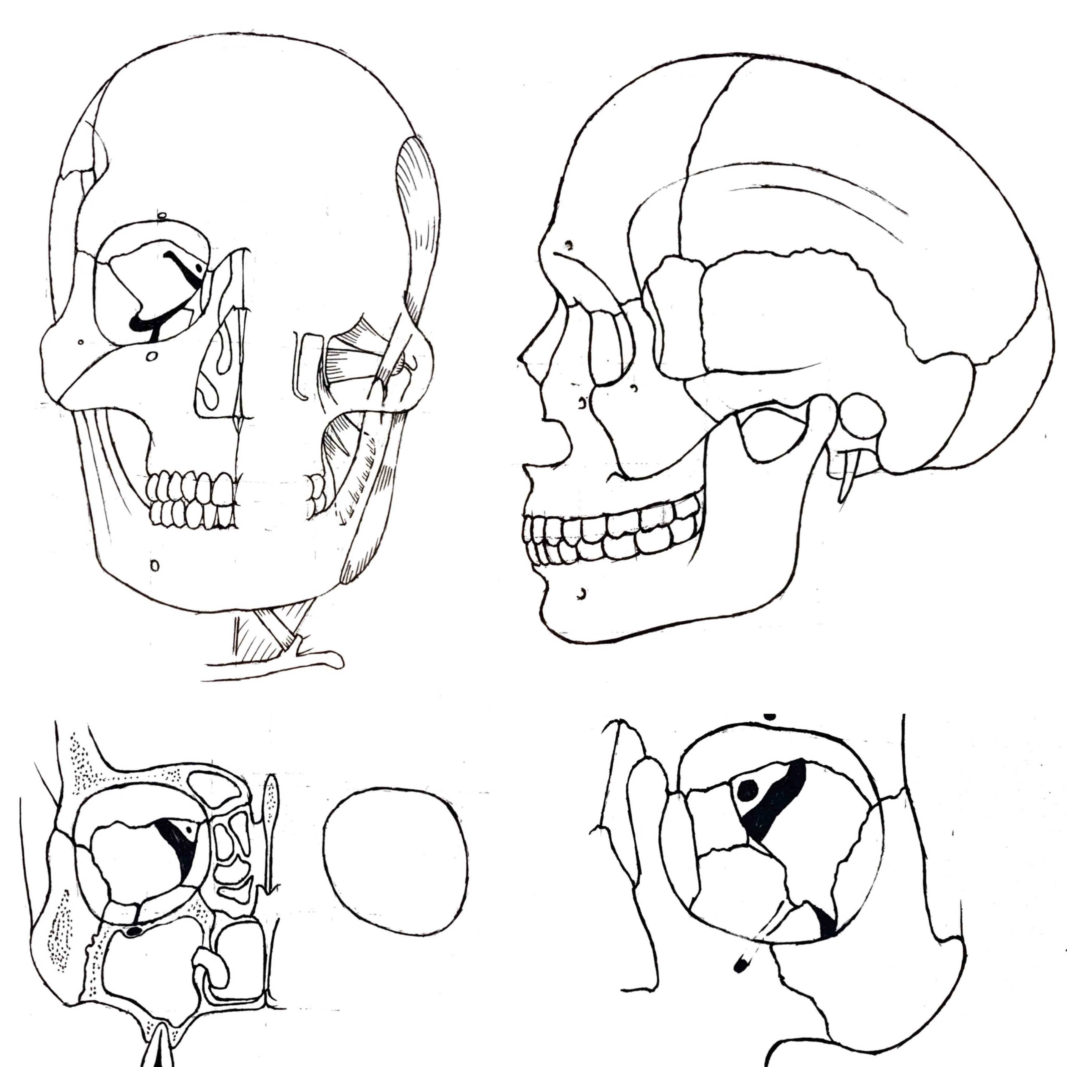
Figure 1: Facial skeleton (clockwise from top left): Facial skeleton with muscles of mastication;
lateral view of facial skeleton; skeletal structure of the orbit; coronal view of paranasal sinuses.
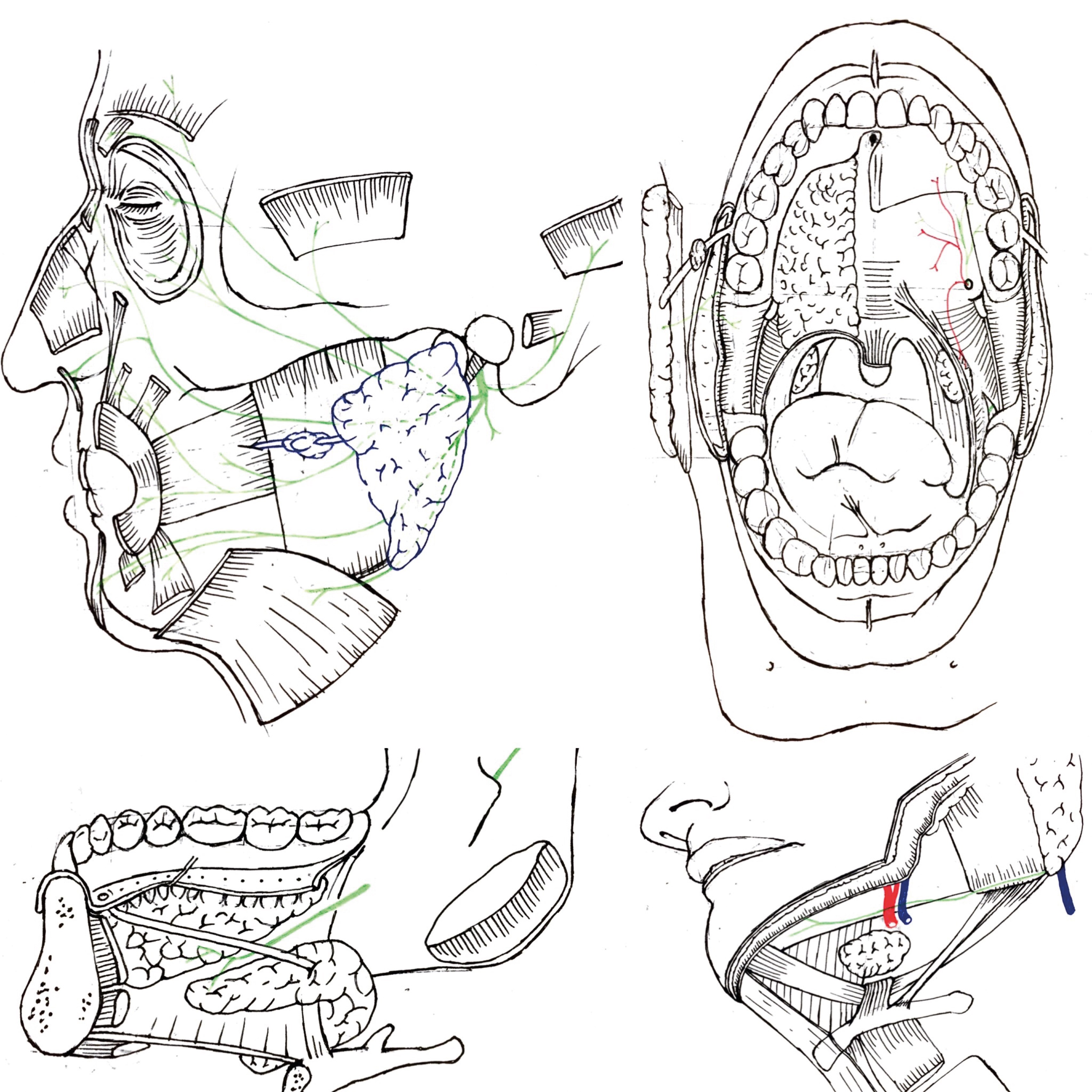
Figure 2: Facial nerve, oral cavity and sublingual / submandibular region (clockwise from top left): Facial nerve and muscles of facial expression, with parotid gland and duct; oral cavity demonstrating, amongst other structures, the palatal and pharyngeal arches; submandibular region with submandibular gland and its relationship with mylohyoid; sublingual region with relationship between submandibular duct and lingual nerve.
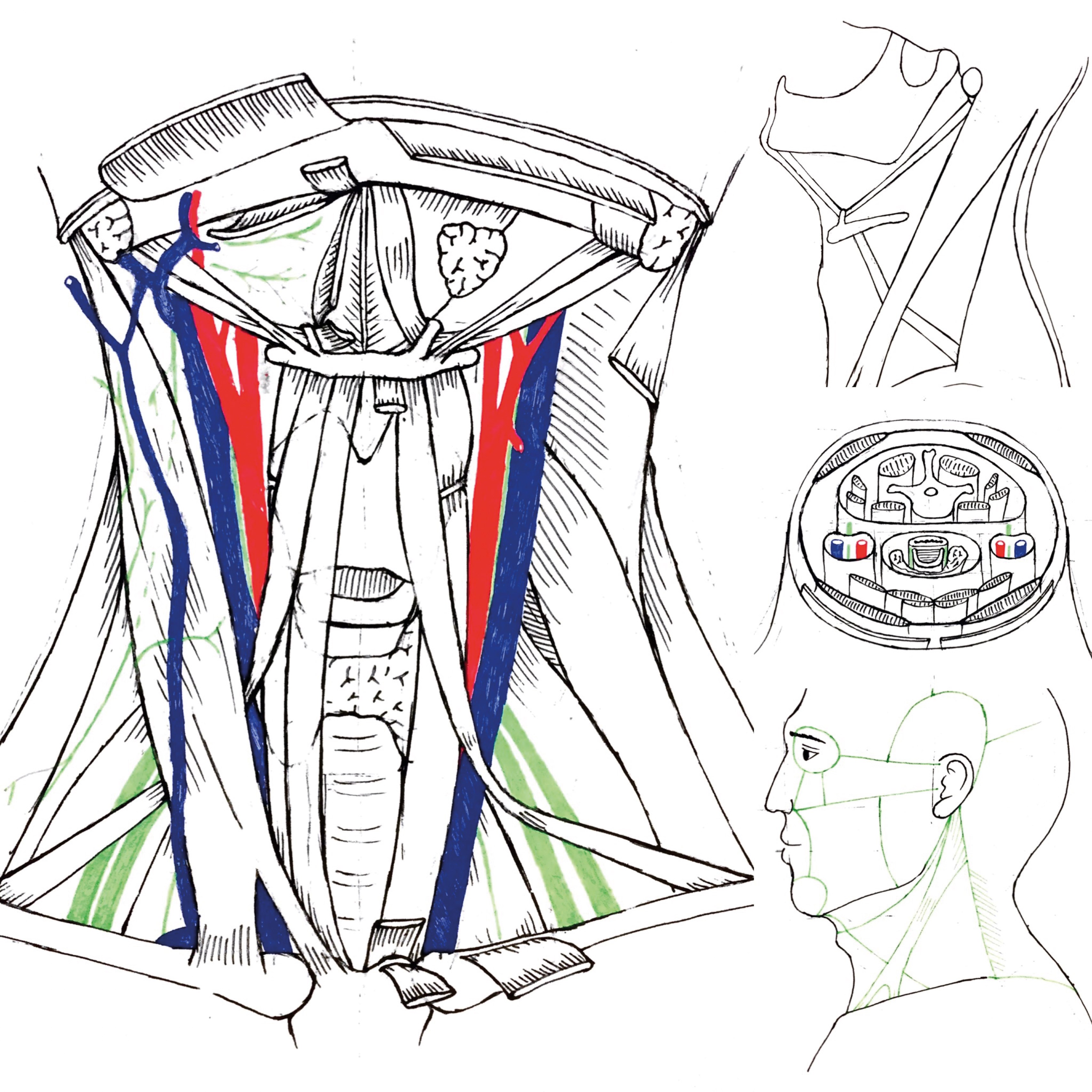
Figure 3: Neck compartments (clockwise from top left): Anterior view of neck showing relationship between the great vessels and strap muscles and sternocleidomastoid muscle; simplified diagram of the anterior and posterior compartments, including sub-compartments; horizontal section of the important fascial layer and compartments of the neck; superficial diagram of the neck compartment divisions.
We introduced several techniques to the students. The first of these was line work, whether in pencil or a pen, and to emphasise that in the context of medical illustration, these should be clean and well-defined, rather than faint or ‘rough’. Additionally, we taught that the crossing of two lines must also be clearly defined and not overlap, i.e. one must be clear of the separation between anatomical structures. Secondly, we demonstrated layers. One can either adopt a lighter shade for underlying structures, or create a cut or retraction of the superior structure in the drawing, which is more technically challenging but can produce a clearer representation. Using colour is also an option, but we felt that students should acquire competence and confidence in simple black and white first and foremost. Our colours of choice for demonstrating purposes were the standard red for arteries, blue for veins and green for nerves. Thirdly, we discussed selecting the medium that the artist felt was most suitable for themselves, i.e. pen, pencil and type of paper, as this selection could subsequently impact on the comfort level of the artist in creating the drawing, enhance the quality of the drawing itself and thus positively impact on the motivation to do more. Our personal preferences were black pens and thick, cream-coloured paper. We also taught that the focus of the session was not to reproduce life-like anatomical illustrations, but rather, to aim for clarity that can translate into better understanding of the anatomical structures and their relations. Finally, our message was to appreciate the relevance of every line in each illustration and its relevance, for it is this last point that we feel underlines how drawing can boost retention of one’s anatomical knowledge.
Overall, the feedback after the session was very positive: the students expressed their enjoyment of drawing anatomy, and felt that they had gained a substantial amount of knowledge in a relatively short span of time and in a unique fashion. The students stated that after seeing the deconstruction of head and neck anatomy through drawing, the subsequent cadaveric demonstration was better understood when compared to before (i.e. without prior drawing demonstrations and practice). A pre- and post-assessment was conducted and we are in the process of formally completing the data analysis.
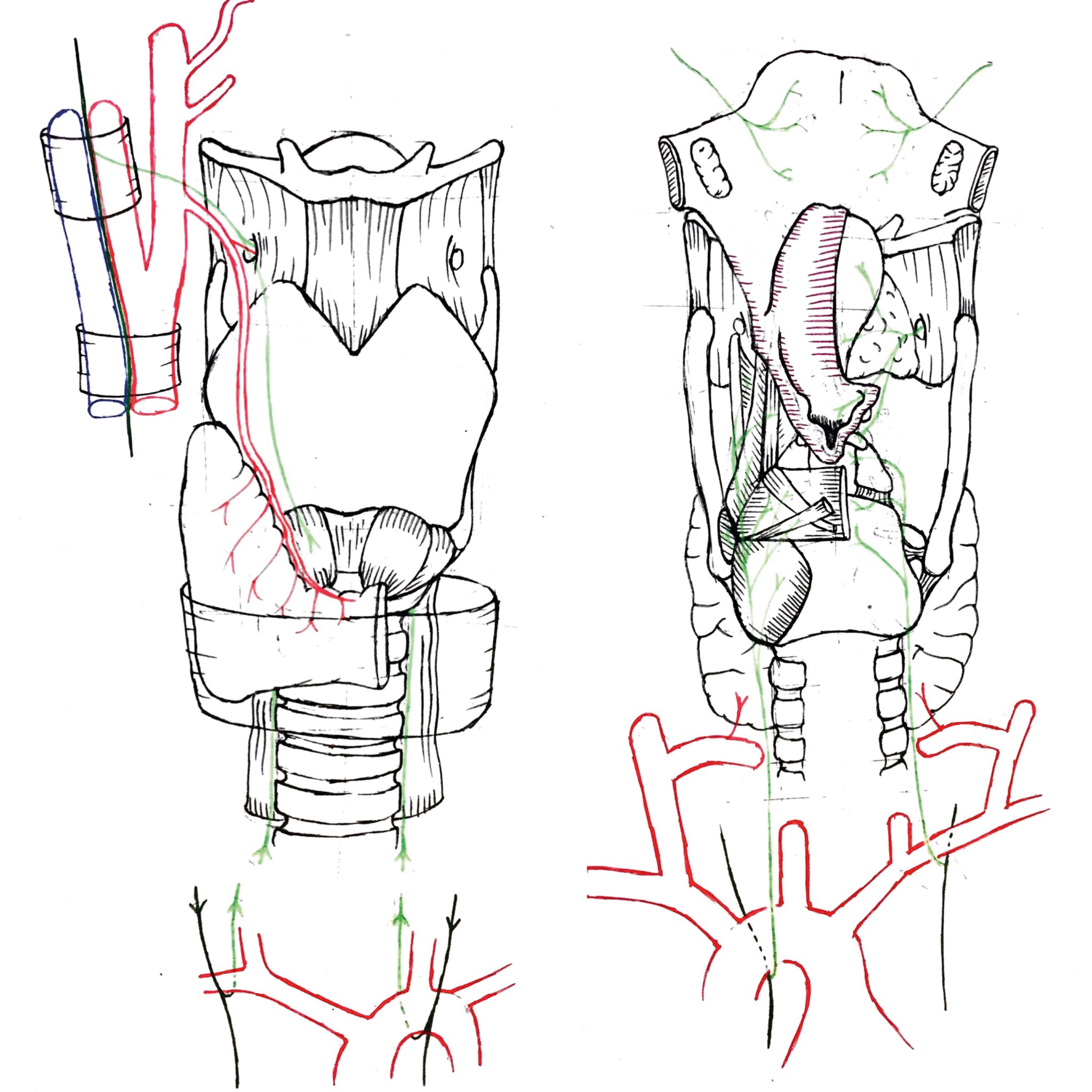
Figure 4: Larynx: (left) anterior view of the larynx and its nerve supply from the superior laryngeal and recurrent laryngeal nerves and its relationship with the thyroid gland; (right) inner view of the larynx demonstrating the epiglottis, glottis, and intrinsic muscles of the larynx and their corresponding nerve supply.
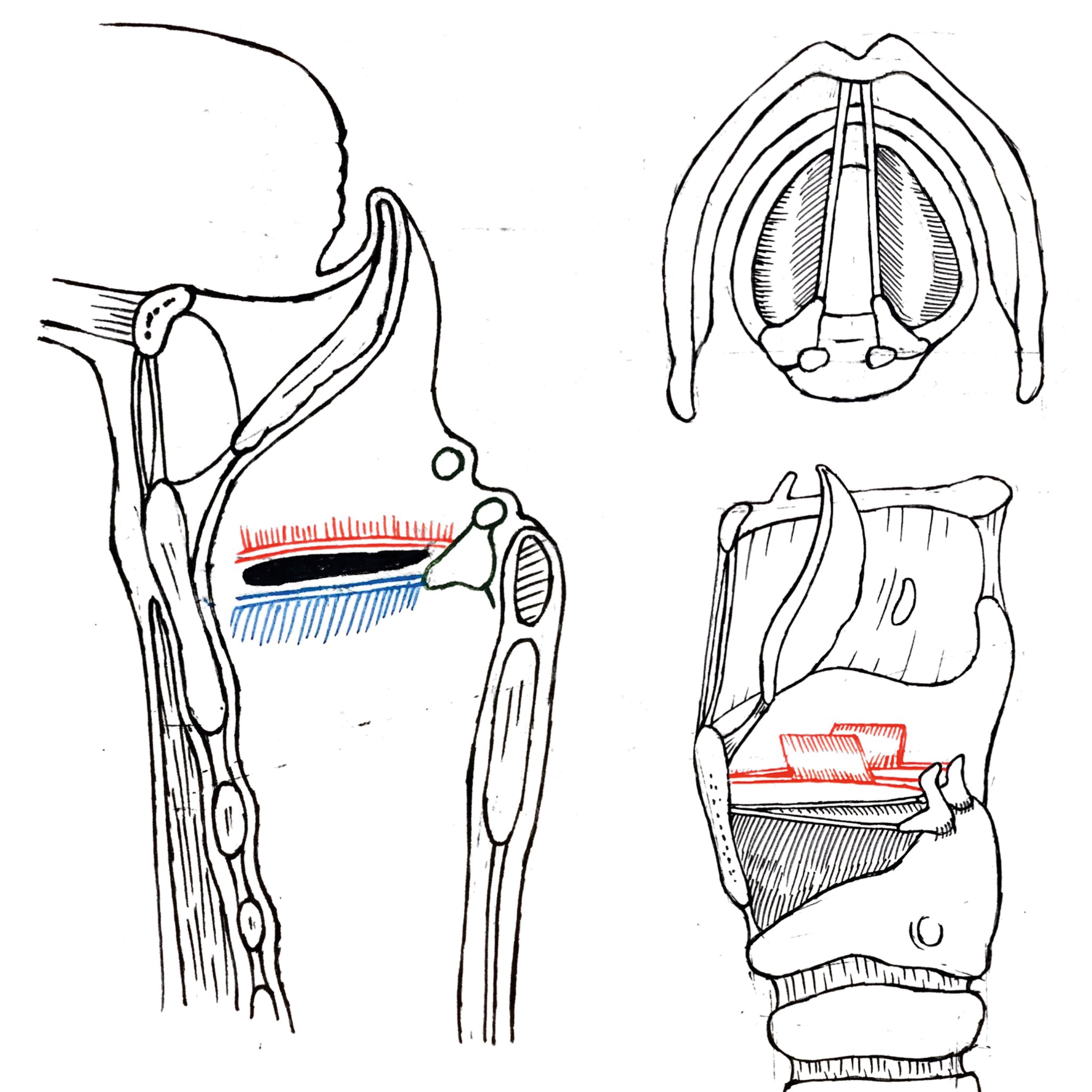
Figure 5: Vocal cords: (left) sagittal view of the vocal cords showing the junction (ventricle) between true (blue) and false (red) vocal folds; (top right) superior view of the make up of the vocal cord from attachments from the arytenoid cartilage to the thyroid cartilage; (bottom right) lateral profile of the larynx and vocal cords, epiglottis and cricoid cartilage and first tracheal ring.
Conclusion
It is evident that art and anatomy have had a mutually reinforcing relationship over the centuries. However, with the teaching of anatomy in the medical school curriculum now shrinking nationwide, and the advent of modern technology and emphasis on textbook learning for exam purposes, the role of art in anatomy teaching is also in danger of becoming less relevant.
Drawing helps students to engage with the complex anatomical planes, thereby allowing them to approach cadaveric dissection with sufficient prior knowledge and to subsequently consolidate it effectively. Students do not require any previous experience of drawing and an individualised approach to teaching anatomy through art is therefore imperative. Also, the emphasis must be on simple line work and anatomical planes, rather than creating life-like or aesthetically pleasing illustrations. To maximise efficiency and ensure adequate and thorough coverage of all topics, the tool of drawing must be used in conjunction with other modalities, i.e. prosections, computer-assisted learning, and lecture-based learning.
We must of course acknowledge that there are also several limitations to teaching anatomy through drawing, the first of these being that not all anatomy tutors can confidently draw to a suitable level for it to be an effective tool in teaching. Secondly, it can be more time-consuming to teach through art demonstrations compared with a lecture-based teaching programme. Thirdly, some concepts are challenging to illustrate, e.g. the nervous system, and other modalities of teaching must be used in these cases.
We intend to publish our quantitative data, to demonstrate whether students had a statistical improvement in their head and neck anatomy knowledge after these drawing sessions. We do hope that studies such as ours will provide much needed evidence of the value of art in teaching anatomy, and that this may find a place in future medical school curriculums, as it already does so in France. It is hoped that in the future these initial teaching workshops may be expanded to help tutors develop their skills to confidently draw anatomy for the purposes of teaching, thus continuing a long historical tradition that is both rich and incredibly satisfying.
References
1. BBC. The Beauty of Anatomy BBC Four; 2014. www.bbc.co.uk/programmes/b04dq8j7 Last accessed April 2017.
2. Clavert P, Bouchaib J, Duparc F, Kahn JL. A plea for the use of drawing in human anatomy teaching. Surg Radiol Anat 2012;34(8):787-9.
3. Jaunoo SS, King TR, Baker RF, Adams HL. A national survey of reasons why students and junior doctors choose not to pursue a career in surgery. Bulletin of The Royal College of Surgeons of England 2014;96(6):192-4.
Declaration of competing interests: None declared.
Acknowledgements
The authors would like to thank to Ms Mimi Borrelli (medical student), Mr Michael Morgan (medical student) and Dr Alistair Hunter (senior lecturer) for their help in organising and delivering our teaching course. Illustrations by Mr Billy Leung.
COMMENTS ARE WELCOME