Uneven tone and pigmentation disorders are frequent problems that motivate patients to seek help. The gold standard for treating the majority of these hyperpigmentation changes is the use of a picosecond laser, as the effect is predominantly photomechanical and photochemical, thus optimal results with almost no downtime and side-effects can be achieved.
Medical management
The first step is to diagnose the pigmentation disorder; dermoscopy and standardised photos with UV analysis should be performed. On patients with diffuse patterns of pigmentation, the skin should be preconditioned for at least three weeks with soft depigmentation agents (e.g. kojic acid 4%), chemoexfoliant actives (e.g. AHA 10%) and sunscreen.
In the case of melasma, patient education and medical care is the cornerstone of the treatment. Beside the behavioural intervention, a full topical regimen should be prescribed, and the laser treatment should only be initiated after four to eight weeks of patient compliance. This regimen brings together photoprotection (UVA/UVB/IR/HEV) and depigmentation, antioxidant, anti-inflammatory and chemoexfoliating agents and tranexamic acid. One should consider adding oral tranexamic acid and P. leucotomos when suitable.
Isolated pigmented lesions
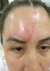
For lentigos, we use the new 730nm Picoway handpiece. The precise wavelength of the PicoWay 730nm handpiece efficiently targets pigment while minimising risk of hypo- or hyperpigmentation and impact on surrounding tissues [1,2]. The handpiece has the shortest pulse duration – 250 picoseconds* – to reduce thermal impact and boost the photoacoustic effect [3]. After thoroughly cleaning the area to be treated, we use the 4mm spot, with fluences ranging between 0.8-1.0J/cm2, covering all lesions. The endpoint is the darkening of the lesion with a perilesional erythema. Sometimes a slight frosting effect may be observed.
For a complete clearance of the lentigo, one to two sessions, minimum four weeks apart are needed. As aftercare, we recommend topical hydrocortisone for five days and sunscreen. The usual evolution is the lesion fading in 10-14 days.
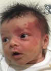
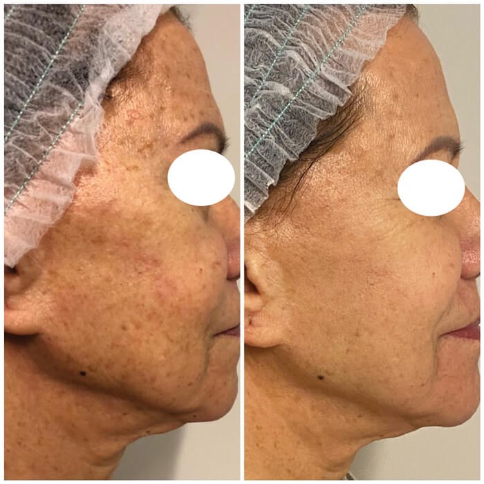
Diffuse pigmentation
For diffuse pigmentation, we start treating the bigger lesions as described before. Afterwards, we use the 532nm Resolve Fusion fractional handpiece to treat the area, with two passes crosshatch. This handpiece provides the picosecond treatment that builds new collagen and elastin [4-6] and blended light technology, reaching more skin surface area. The short pulse duration (375ps) creates more photoacoustic and less thermal damage [3]. The usual energy setup is 0.35-0.50mj/px at 7Hz, it is required about 3500-4000 shots to cover the whole face. After the laser treatment, we apply a soothing mask to improve patient comfort. The aftercare is similar as described before. The typical post procedure is a mild redness that lasts up to five days. The pigmented lesions will darken, form a very thin crust and scab after about seven days. For a complete treatment, two to three sessions are needed, at a minimum interval of four weeks.
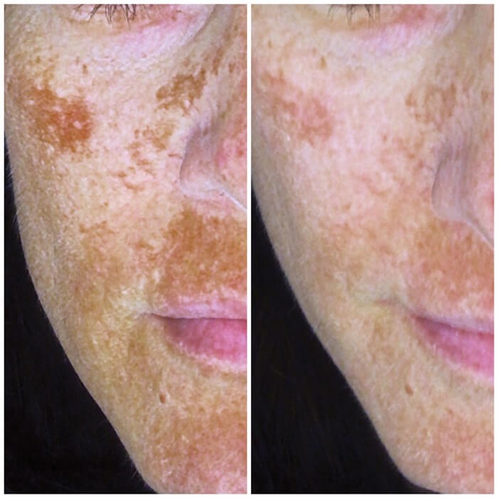
Melasma
Laser therapy is an important adjunctive resource to deal with dermal pigmentation deposits and the vascular component. The first step is to control the vascular component, using the Vbeam Prima at 595nm (15mm, 4.00-4.50J/cm2, 3ms, DCD 30/20). Following this, we perform a melanocyte dendridectomy, using the Picoway Zoom handpiece, 8mm spotsize, 0.60-0.75J/cm2, doing two general passes and an extra double pass on the pigmented area.
Then, we target the melanosome again and perform a fibroblast stimulation using the 1064 Resolve handpiece, with an energy of 1.3-1.5mJ/px, full face double pass along with an extra double pass on the pigmented areas. This increases the skin permeability, allowing laser drug delivery with tranexamic acid. Typically, a mild erythema lasting up to 48 hours is observed. As aftercare, we recommend topical hydrocortisone for five days and sunscreen. The number of sessions is variable, but the average is four. The recommended minimum interval between treatments is four weeks.
Conclusions
The Picoway and its picosecond technology is the front end for treating pigmentation disorders efficiently. The use of holographic fractionated handpieces expands the traditional use of these types of lasers, with its regenerative (thus rejuvenation) capabilities.
References
* Based on published manufacturer specifications at the date of publication. Candela – Data on File, 2020.
1. Artzi O, Mehrabi JN, Koren A, et al. Picosecond 532-nm neodymium-doped yttrium aluminium garnet laser-a novel and promising modality for the treatment of café-au-lait macules. Lasers Med Sci 2018;33(4):693-7.
2. Serup J, Bäumler W (eds): Diagnosis and Therapy of Tattoo Complications. With Atlas of Illustrative Cases. Karger: Basel; 2017;52:113-23.
3. Kung KY, Shek SY, Yeung CK, Chan HH. Evaluation of the safety and efficacy of the dual wavelength picosecond laser for the treatment of benign pigmented lesions in Asians. Lasers Surg Med 2019;51(1):14-22.
4. Brauer JA, Kazlouskaya V, Alabdulrazzaq H, et al. Use of a picosecond pulse duration laser with specialized optic for treatment of facial acne scarring. JAMA Dermatol 2015;151(3):278-84.
5. Tanghetti EA, Tartar DM. Comparison of the cutaneous thermal signatures over twenty-four hours with a picosecond alexandrite laser using a flat or fractional optic. J Drugs Dermatol 2016;15(11):1347-52.
6. Tanghetti EA. The histology of skin treated with a picosecond alexandrite laser and a fractional lens array. Lasers Surg Med 2016;48(7):646-52.
Declaration of competing interests: The author has been reimbursed by Candela Medical for speaking at congresses and training sessions.
COMMENTS ARE WELCOME