Ageing related weakening of the inferior orbital septum and increase of orbital fat volume is considered to result in lower lid prominence. Attenuation of the septum allows the gradual herniation of one or more of the three intraorbital fat pads. Furthermore, gravitational descent of the mid-face aggravates the fat protrusion of the lower eyelids. Jo and others [1] in 2017 classified 1034 Korean eyebag patients (269 men, 765 women) using their frontal photos into medial bulging (M) type (17%), medial and central bulging (MC) type (49%) and medial to lateral bulging (MCL) type (34%).
In our practice the mainstay of treatment of lower eyelid bulging arising from orbital fat protrusion is surgery by lower blepharoplasty which is carried out by a transconjunctival approach assisted by carbon dioxide laser [2]. This approach is applicable to nearly all cases as it prevents less oedema and scarring to the orbicularis muscle and possibly reduces ectropion in patients with decreased lower eyelid tone. A lower rate of complications has been observed by the transconjunctival group with greater patient satisfaction [3].
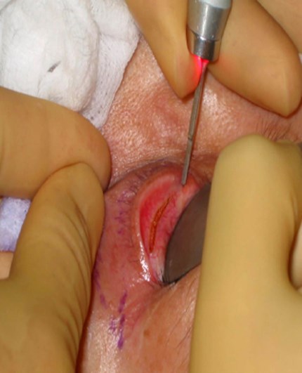
Figure 1.
Transconjunctival lower blepharoplasty is usually done under local anaesthesia supplemented by oral sedation and antibiotics prophylaxis. After conjunctival incision by carbon dioxide laser in the cutaneous wave mode (Figure 1), the preseptal plane is dissected to reach the septum behind the orbicularis oculi muscle. A small Desmares retractor is used to retract the lower eyelid and to search for the fat pads. After incising the septum, the central fat is identified first. The blood vessels associated with each fat pad have to be cauterised with a separate diathermy instrument for complete haemostasis (Figure 2).
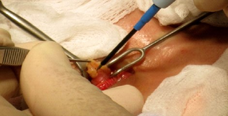
Figure 2.
The small Desmares retractor is replaced with a large Desmares retractor which is burnished or dulled to avoid reflection of laser light. After securing the Desmares retractor flush with the orbital rim, the central fat pad is lifted up against the Desmares retractor and the focused laser beam is used to excise the protruding portion of the fat pad (Figure 3).
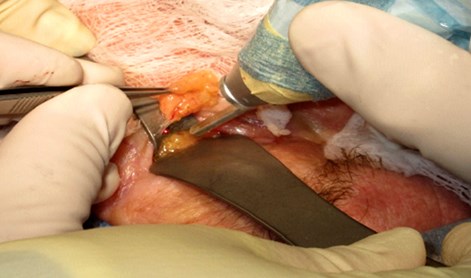
Figure 3.
CO2 laser provides simultaneous haemostasis especially when applied in the defocused mode. With appropriate retraction, the medial fat pads and the lateral fat pads are next searched, identified and appropriately excised. The medial fat pad is harder to expose and identify, it is also bounded laterally by the inferior oblique muscle which must be preserved. The endpoint is to remove an appropriate amount of fat down to and flush with the inferior orbital rim. Experience is required in removing the correct amount of fat pad in order to minimise residual fat bulging and avoiding an empty socket look due to excessive fat removal (Figure 4).
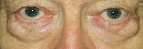
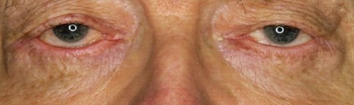
Figure 4.
Usually, the conjunctival incision is approximated underneath the conjunctiva with an absorbable 6/0 vicryl suture to facilitate proper healing. For patients with excessive lower eyelid skin, especially in patients aged 55 or above, simultaneous excision of pinched skin (about 2-3mm width of skin) is carried out. The skin defect after haemostasis is usually sutured with interrupted and continuous 6/0 nylon sutures. This combined inside and outside approach minimises trauma to the orbicularis muscle. Topical antibiotic ointment, artificial tears and antibiotic eye drops are prescribed for after care along with cold compresses for the first 36 to 48 hours.
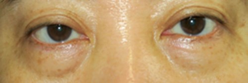
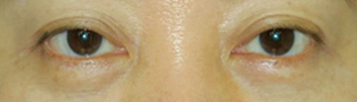
Figure 5.
Patients should be informed of the possible side-effects and complications after lower blepharoplasty: ecchymosis, oedema, bleeding from conjunctival incision, orbital haematoma which may cause blindness, temporary visual disturbances from conjunctival swelling and bleeding, entropion, ectropion, diplopia, dry eyes, infection, residual bulging, uneven surface as well as an increase in dark circles, wrinkles or depression from excessive fat removal. Follow-up for the first six months is required to reach final clinical outcome (Figures 4 and 5).
References
1. Jo SJ, Kim HS, Park JT, et al. Assessment of age and sex related changes in baggy lower eyelids using a novel objective image analysis method: orbital gray scale analysis. J Cosmet Dermatol 2018;17:874-80.
2. Chung ST, Rho NK. Surgical and non-surgical treatment of the lower eyelid fat bulging using lasers and the energy-based devices. Med Laser 2017;6(2):58-66.
3. Rancati A, Jacovella P, Zampieri AE, et al. Lower blepharoplasty review, transconjunctival vs transcutaneous approach. Modern Plastic Surgery 2015;5:1-8.
Declaration of competing interests: None declared.
COMMENTS ARE WELCOME