A pre-tibial laceration is a common and serious wound, particularly in the elderly where co-morbidity from smoking, peripheral vascular disease, diabetes, malnutrition and tissue-paper-thin skin are major influencers of outcome. Degloving injuries may be seen as preterminal events in the frail patient after the most minor trauma.
With such a variety of pre-morbid conditions, degree of tissue trauma and management protocols it is little wonder that outcomes vary and most, if not all, published studies are full of confounders such that meta-analysis is meaningless.
There are a number of classifications that have been suggested to try and compartmentalise good practice, the best being described by Dunkin, et al. in 2003 [1] but in reality the outcome depends upon the starting point of health, the pre-morbid quality of the tissues, the degree of crush and shear of the tissue planes, but probably, and most importantly, the management of the wound and rehabilitation.
Pre-tibial lacerations often exhibit delay in healing over months. Skin coverage of a debrided pretibial wound may involve relocation of degloved skin flaps and or skin graft. The wounds may be left to heal by secondary intention, but where there is significant skin loss this may take months. The former may be sutured, steri-stripped or closed with a combination of both – or even suturing through steri-strips to redistribute tension away from thin friable skin edges.
When wounds break down, delayed wound healing is inevitable. To speed up wound healing by secondary intention, lasers and focused extracorporeal shockwave therapy (fESWT) have been proven to be effective.
The first few weeks of care need teamwork and a regular care plan devised by specialist practitioners. The primary intent is to quickly resolve any developing infection and encourage healing and repair in a way that may avoid further surgery. The secondary intent is to encourage early mobility and to minimise scar formation.
Lower-limb skin heals well when sutured, provided the skin edges have a healthy blood supply and are approximated with little tension. There must be no dead space behind the traumatised skin flaps or seroma / haematoma will cause necrosis, but also too much external pressure on the skin will also cause flap necrosis. Promoting blood vessel ingrowth and vascular flow is integral to rapid wound healing. Thereafter a proactive rehabilitation programme is vital for the individual to return to a pre-injury state.
Adjuncts to wound care were available to clinicians even in the 1980s – for example, the ‘Space Laser’ was used in 1988 for speeding up healing of open palm wounds after Dupuytren’s contracture excision in hand surgery. These wounds healed one week faster using the space laser. Controversially at that time there was a transition to immediate mobilisation after soft tissue sports injuries, such as acute tendoachilles repair, to get elite athletes back to compete. This principle was also applied to mobilise extensively injured burn patients, even those on ventilators.
Early mobilisation encourages revascularisation and tissue plane mobility and better outcomes. Negative pressure vacuum devices or use of the open skin graft exposure method, whilst giving excellent results, will require bedrest and immobility and in the elderly this approach can be fatal. This case report demonstrates the beneficial outcome of combining fESWT and supervised immediate mobilisation in a patient with a significant pretibial wound. It is relevant when considering adjuncts to care that speed up healing and give the best chance for return of function from most wounds.
Case history
The patient is a fit and healthy, non-smoking, non-diabetic 62-year-old man with no significant medical history. On 4 October 2024 he fell through a hatch in the floor of his luxury yacht whilst sailing off the coast of Ibiza, Spain. He was airlifted to hospital on the mainland with an extensive pretibial crush type soft tissue flap injury on his left lower leg, which was debrided and primarily closed. He was given antibiotics for 10 days and discharged.

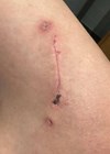
Figure 1: The initial wound on the left anterior shin presenting four days after injury. The wound has advancing cellulitis and shows full thickness skin necrosis and an inflamed / infected wound line. After surface cleaning and superficial debridement, the wound was treated with fESWT. This continued twice weekly until healed.

Figure 2: Preparation for treatment.

Figure 3: The ESWT unit used to treat the patient (STORZ MEDICAL).
On 8 October 2024, four days after injury, he presented to the first author at his London Practice and the wound is shown in Figure 1. There is extensive full-thickness necrosis of the central wound area and an infected necrotic suture line with advancing cellulitis. The wound was cleaned, and he was given antibiotics. The sutures were removed at two weeks. At this first presentation, a course of fESWT (Stortz R) commenced (Figure 2 and 3). Treatment was topically delivered twice weekly using the handheld device with sterile contact gel as lubricant. The gel was applied directly over the healing area at settings of 0.02–0.03mJ/mm2 equating to 1500 shocks at 4Hz. He was encouraged to mobilise on crutches for two weeks and was thereafter full weight bearing and walking distances twice daily with a tubigrip support over a wound covering of non-adherent dressing. Between times he would elevate the leg at home. After eight weeks he was back in the gym training, adhering to a unique lower limb rehabilitation programme developed by the first author. He was also given advice on sun exposure months. He dispensed with his support tubigrip after six weeks.

Figure 4: The wound after 22 days showing reduced inflammatory change and shrinkage of the crushed tissues.

Figure 5: The wound at 31 days with significant shrinking of area and manageable dry crusting.
The edges of crusting have been trimmed as they raise from the healing area.

Figure 6: The wound at 35 days. All that remains is a thin dry crust.
The patient is now fully ambulant and is fully healed after natural eschar separation a few days later.

Figure 7: The residual scar. Note the smoothness of skin and the amount of wound contraction with hair appearing.
The skin is sensate with no hypersensitivity.
The chronology of the healing wound is shown in Figures 4–7. The wound had fully healed within 35 days and he was discharged. Seven months later the patient presented in clinic for unrelated neck physiotherapy and wished to share his comments: “The skin at the site of leg injury is smooth and soft with no evidence of a scar other than what now looks like a fine line tattoo on my foreleg, with no discomfort to touch.”
Summary
Today, there are opportunities to consider other modern adjuncts that may improve the duration and quality of wound healing. Although restriction of opportunity in the NHS will be related to availability of staff and the purchasing of costly advanced technology, there is much evidence that this can be a cost-reducing and highly effective way of managing very difficult wounds. Immediate mobilisation reduces inpatient dependency and likely gives a better functional and aesthetic outcome, and requires less secondary or revisional surgery.
Quite how these technologies work is under investigation, but a likely pathway involves the hyper-stimulation of healthy cells in the vicinity of the wound. This triggers an exosomal discharge from these cells which orchestrates other cells to start their healing activities. The evidence for their involvement in wound healing is strong [2,3]. Exosomes contain signal proteins and chemicals that will promote neovascularisation, fibroblast migration and collagen proliferation. Exosomes are also a focus of attention in regenerative and anti-ageing medicine.
Exosomes are small membrane-bound vesicles expelled by cells in response to a need, by stimulation or trauma. They transmit inter-cellular messages to other cells that cascade to promote healing through capillary ingrowth, fibroblast proliferation, collagen regeneration, reduce swelling and increase blood flow likely through the vasodilatory effect of nitric oxide. K lasers and shockwave therapy are known to promote wound healing and were both available for this patient, but after discussion with the patient, the first author elected to use a course of fESWT with successful outcome.
We encourage others to consider adjuncts to wound care that benefit patients by promoting rapid wound healing and physical recovery and lessen the cost to patient care.
References
1. Dunkin CS, Elfleet D, Ling C, et al. A step-by-step guide to classifying and managing pretibial injuries. J Wound Care 2003;12(3):109–11.
2. Crevenna R, Mickel M, Schuhfried O. Focused Extracorporeal Shockwave Therapy in Physical Medicine and Rehabilitation. Curr Phys Med Rehabil Rep 2021;9:1–10.
3. Pölzl L, Nägele F, Hirsch J, et al. Exosome Isolation after in vitro Shock Wave Therapy. J Vis Exp 2020;163:doi:10.3791/61508.
Declaration of competing interests: None declared.