A youthful dorsal hand is characterised by a supple, smooth skin texture and contour, with minimal prominence of the dorsal veins. Over time, extrinsic (e.g. sun damage and smoking) and intrinsic (bone remodelling and soft tissue volume changes owing to ageing and disease processes such as arthritis) result in textural changes, dermal atrophy, prominence of the dermal veins, visibility of the tendons, soft tissue volume loss, and bone and joint changes.
These changes can be significantly improved with the aid of volumising agents such as calcium hydroxylapatite or hyaluronic acid dermal fillers. Effective modalities described in the literature to improve texture and pigment changes can include chemical peels, photodynamic therapy, intense pulsed light, as well as lasers.
There are key anatomical concepts regarding the anatomy of the dorsum of the hand, which includes the skin, soft tissue, areolar tissue / dorsal laminae, as well as intrinsic muscle activity and wasting. The superficial layer is the first and is located between the dermis and the dorsal superficial fascia which does not contain structures and may be adherent to the deep dermis, in an aged hand. The intermediate fat layer resides between the dorsal superficial fascia and the dorsal intermediate fascia, the latter is an extension of the antebrachial fascia of the forearm. The intermediate layer contains the dorsal veins and sensory nerves. The deepest and final fat plane is located between the dorsal intermediate fascia and the dorsal deep fascia, which covers the interosseous muscles and the metacarpal bones.
The extensor tendons are located between the dorsal intermediate fascia and the dorsal deep fascia. There is potential communication between the palmar and dorsal vessels through perforators, which pass through the interosseous spaces, which lead to the end arteries in the digits. There is potential for material injected in the dorsum to pass by a retrograde mechanism into the palmar circulation and enter the end artery of the digits. An assessment of the motor and sensory activity of the palmar region of the median, radial and ulnar nerves, should be performed. In addition, the arterial supply should also be assessed by utilising the Allen test.
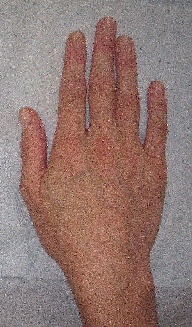
Figure : Before augmentation using dermal fillers.
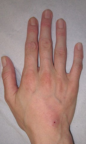
Figure 2: After augmentation using dermal fillers.
A detailed understanding of the anatomy will enable practitioners to place products accurately, safely and minimising the risk of any complications (e.g. bruising). Essential components to consider include the skin, soft tissues (areolar tissue / dorsal laminae), and intrinsic muscle activity and wasting. It is also crucial that any veins and tendons are identified to avoid traumatic injury. The use of validated aesthetic scales are a valuable consultation tool to assess the pre-treatment presentation and the predicted degree of correction. This will enable patients and practitioners to discuss the realistic expectations regarding the possible outcomes of treatment.
In line with all dermal filler treatments, a strict aseptic technique is imperative in light of the close communication of the compartmental spaces of the hand; the patient’s hands are washed with an alcohol based solution and the dorsum cleaned with chlorhexidine. Current literature suggests that a safe plane for product placement is between the dermis and fascial layer, using either a 22G or 25G, 38mm or 50mm cannula approach, creating a subdermal space for injection. With appropriate use, a cannula may be less likely to perforate vessels to ensure product remains in the appropriate plane. An ideal injectable substance to contour the dorsum of the hand should be durable to withstand considerable dynamic demand, as well as pliable with significant longevity. My preferred product choice for dorsal hand augmentation is calcium hydroxylapatite, which is now available pre-mixed with lidocaine and also has FDA approval for dorsal hand rejuvenation.
Retrograde threads of product (0.5-0.1ml per pass) placed through a proximal insertion point can be placed to safely correct many of the age-related changes. Current literature suggests a safe plane for product placement is between the dermis and fascial layer. After correction, the product is massaged gently to ensure even distribution and the patient is advised to keep their hands clean and dry following treatment, to avoid wearing any creams and abstaining from vigorous activity for 48 hours.
It is crucial that practitioners are up to date on their anatomical knowledge before treating the hands with any injectable products. Clinical anatomical studies continue to emerge, analysing the effectiveness of different modalities, including combination treatments. Awareness of the dorsal hand anatomy and vascular system will ultimately increase the degree of accuracy in product placement, and decrease the risk of complications.
COMMENTS ARE WELCOME