Breast cancer has become so common that most people reading this article will know someone (either professionally or personally) who has been affected by breast cancer. One of the most common treatments for breast cancer is removal of the ‘whole’ breast (mastectomy).
In various parts of the developing world it is still a common practice for women who have undergone a mastectomy to fill out their bra with cotton wool or other materials. Some women will use various kinds of external prosthesis to mask the fact that they have had a mastectomy. (Editor’s note: for more information on the external prosthesis please refer to the following article http://www.medscape.com/viewarticle/460148). The external prosthesis is commonly used as a temporary measure in women who are seeking a delayed reconstruction. It is unfortunate that in a global context there is a widespread ignorance of the reconstructive options amongst patients, which also extends into the medical community. Fear of the process of breast reconstruction as well as the costs of surgical reconstruction are further considerations that need to be recognised and addressed.
There is a different and more positive story in the more developed parts of the world. In the United Kingdom, there have been increasing numbers of immediate and delayed post mastectomy patients requesting and receiving reconstruction. This is partly due to the very significant improvement in traditional reconstructive techniques, the advent of new ones and also partly due to increased awareness of the nature and availability of reconstructive options. The education of the public, particularly through online articles and features has created a far more knowledgeable patient community. The internet has also created a more enlightened medical fraternity, especially in allied specialties. The combined result has been a very positive advance in the support for patients in their quest to try and restore an acceptable degree of normalcy to their appearance and ultimately their function.
Apart from the obvious concerns about the underlying malignancy and fear of recurrence, a woman who has had a mastectomy may encounter a number of other issues. These range from the physical absence of the breast mound and a feeling of loss of femininity, self-consciousness or anxiety in clothing, self-consciousness due to wearing the external prosthesis, particularly as these may be displaced or even fall out of the bra causing embarrassment, a negative body image, loss of libido or sexual interest, depression and mood disturbances. For patients who have a mental health issue, mastectomy might actually tip them over into a significant crisis. It is clearly evident that there is a need to reconstruct or address this loss of the breast in many patients.
That brings one to ask what breast reconstruction is and how it can help in light of these concerns. In the context of the focus of this article, I see breast reconstruction as a process of creating aesthetically pleasing and symmetrical breasts following removal of one or even both breasts. It is an attempt to restore the breast appearance, not only in volume, size but also in projection. In other words, a well reconstructed breast has to look like a breast and if the mastectomy has been only on one side, then it has to look like the contralateral breast. It should not only feel realistic to the patient but also appear realistic to those around her.
The objective or the aim of breast reconstruction is to help restore the patient’s quality of life. The reconstructed breast mound helps reduce the emotional and psychological disturbances associated with mastectomy. It enables the patient to get back into their pre mastectomy social life more quickly and comfortably and plays a very significant part in the rehabilitation of the patient.
It is reasonable to state that there is no ‘ideal’ form of breast reconstruction, but there is an ideal for each patient. Despite this, there is a gold standard, which has to be reconstruction with autologous tissue.
Consultation
After discussion in the multidisciplinary team meeting, suitable patients for immediate post mastectomy reconstruction are reviewed in the Combined Breast Reconstruction clinic (involving the breast surgeon and plastic surgeon). For patients contemplating immediate reconstruction, this is a very sensitive period as the diagnosis of the cancer is recent, combined with the prospect of losing one or both breasts. One has to decide whether in such circumstances, informed consent of the process is possible for the particular patient. There is a spread of patients, from those who have researched breast reconstruction extensively (even in the short period since diagnosis) with ready questions, to those who appear ‘detached’ and not engaged. One may argue that the latter group are unlikely to be good candidates for immediate reconstruction.
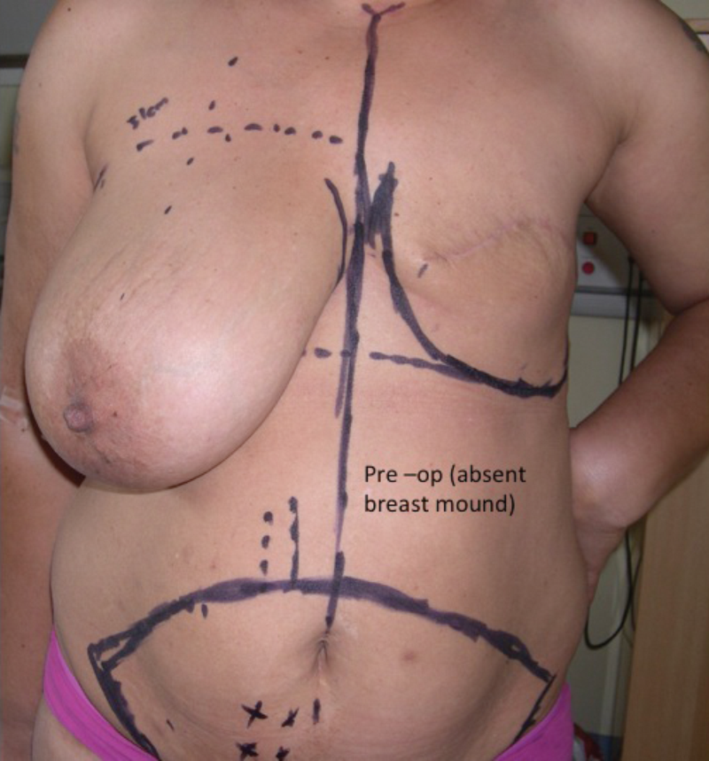
Figure 1: Pre-op (before reconstruction). Patient had left mastectomy.
Those patients presenting for delayed reconstruction are usually referred by the consultant breast surgeon, consultant oncologist and occasionally by their GP and will have had time to think about reconstruction and / or discuss it with one of the referring physicians and other patients.
During the consultation, I enquire about treatments received or planned, such as radiotherapy, chemotherapy and hormonal therapy. These can have an effect intraoperatively (blood vessels friable from radiotherapy, fibrosis and even ‘absent’ blood vessels following previous surgery; increased risk of extrusion of implants) in delayed reconstruction and also affect the result of the reconstruction in immediate reconstruction (radiotherapy shrinking the tissues afterwards, adverse capsular contracture).
Information about co-morbidities, such as diabetes mellitus, hypertension etc., lifestyle habits (smoking), medications including herbal remedies and allergies is essential. This information is necessary to give the patient a balanced view about choice of reconstructive technique and the risks of the surgeries in the presence of such factors.
It is my practice to see the patient more than once before embarking on the process of breast reconstruction. In cases of immediate reconstruction, the interval can be as short as a week. Much information is given and it takes time to assimilate or digest this. Attendance with a close family relative such as a husband or sister or significant other is encouraged because these form part of a good support network.
I give patients a breast reconstruction leaflet for further reading and encourage them to write down their questions on paper, which they can bring to the next consultation. Patients are also provided with information about breast reconstruction awareness meetings. These meetings are run by specialist nurses and attended by previous breast reconstruction patients as well as those considering reconstruction. It offers a valuable opportunity for prospective patients to interact with those who have ‘travelled the path’ and get a patient’s perspective rather than purely a doctor’s information about the process. I find it useful for some patients to review clinical pictures of previous patients. This is always done with a warning that every patient is different and the result or appearance depends to a great extent on the oncological surgery especially the extent of skin excision.
It is hoped that all the above deals with the concerns of the patient, and the patient is therefore able to make a decision and give informed consent to the most appropriate breast reconstructive technique.
Stages
Breast reconstruction is best described as a reconstructive process. It usually occurs in stages. For purposes of our discussion I divide the reconstructive process into three stages:
- Creation or restoration of the breast mound.
- Symmetrisation and creation of nipple.
- Areola reconstruction.
These stages can be combined. In other words they do not necessarily need to take place serially. For example, it is not unusual to do a contralateral breast reduction or lift (symmetrisation) in the first operation. On the other hand, a symmetrisation procedure can be carried out without nipple reconstruction in the reconstructed breast to allow time for the breasts to settle and for the patient to be satisfied with symmetry before the appropriate position of the new nipple is decided.
However, the completion of the process requires tattooing around the nipple, which starts about six weeks after nipple reconstruction.
Stage 1 – restoration or creation of the breast mound
There are broadly three ways of creating and restoring the lost breast mound. The materials can be purely non-autologous materials or purely autologous (patient’s own tissues) or a combination of both.
Non-autologous (tissue expansion)
This is mainly the use of tissue expanders and breast implants. The expanders are usually implanted in a sub-pectoral plane to add another layer of vascularised soft tissue cover. Using the inflation or injection port, the expander is inflated with normal saline starting intraoperatively and continuing postoperatively. This occurs over weeks to months and the skin is stretched gradually until a satisfactory mound is created. The expander is then removed and replaced with a permanent silicone implant.
In recent years the expander implant is more commonly used as they can be left in place as ‘permanent’ implants. Under local anaesthetic, through a very small incision the port is removed leaving the expander implant in place. Plans have to be made to replace the expander implant with a permanent implant at the same time, if there is a malfunction or rupture of the valve system during the disconnection of the port tubing from the main expander implant base. So I warn and consent patients for this and also get the appropriate size of implant ready on the shelf. Also, whilst the procedure is usually performed under local anaesthetic, the patient is starved in case of the need to convert to general anaesthesia if an immediate implant is needed.

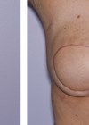
Figure 2: Post-op – left breast mound restored.
The advantages of this technique are:
- Patient’s breast skin is stretched which is the best match with the contralateral breast skin.
- No new scars are introduced as the surgery is carried out through the mastectomy scar.
- A shorter duration of surgery (usually one to two hours).
- A shorter hospital stay (usually one or two days).
- A shorter learning curve than for the other methods.
This should not be misconstrued necessarily as being an easier procedure. There are subtle but significant modifications in technique that make the breast look aesthetically pleasing.
The disadvantages include:
- Pain or discomfort during episodes of expansion.
- Injection port problems such as:
º pain over the injection site
º flipping of the port making inflation difficult or impossible
º disconnection, causing deflation of the implant. - Concerns and problems with implants also exist such as:
º adverse capsular contracture
º wrinkling of implant
º infection and need for explantation and an interval period of three
months or more before re-insertion
º rupture.
Though breast implants have not been shown to constitute any significant health hazard to women, scientifically unsubstantiated claims in the press over the last two decades and especially in the last few years have left concerns in the minds of some patients, especially with respect to cancer causation and detection.
Autologous
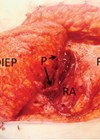
This is regarded as the gold standard. Various donor sites exist. Common sites include the lower abdomen (DIEP – deep inferior epigastric perforator flap, SIEA – superior inferior epigastric artery flap), trunk (ELD – extended lattismus dorsi muscle flap, TAP –thoracordorsal artery perforator flap, intercostal artery perforator flap), buttocks (SGAP, IGAP), and inner thighs (transverse upper gracilis – TUG flap). Other sites include the lower back (LAP – lumbar perforator flap), flanks (Ruben’s flap – deep circumflex iliac artery flap), anterior thighs (anterolateral thigh flap) and posterior upper thigh (profunda artery perforator flap – PAP). Any source of significant vascularised soft tissue is a potential donor site for breast reconstruction material.
The recipient vessels are usually the internal mammary vessels and thoracodorsal vessels. Any other pedicle in the vicinity can also be used as recipient vessels including lateral thoracic, transverse cervical vessels and perforators. It has significant advantages:
- There is possibility of obtaining enough tissue to form a good-sized breast.
- There is no fear of ‘rejection’.
- Complications such as infection, wound dehiscence, partial loss of flap and mastectomy skin flap problems are managed better and often lead to a satisfactory outcome. This is not the case with non-autologous reconstructions.
- The autologous tissue consists of skin and fat which is more similar to the breast tissue in consistency and density than non-autologous tissue. As a result, autologous reconstruction looks and feels more like a normal breast in the long-term, particularly with regard to ptosis; and arguably is more durable than non-autologous reconstruction.
- For certain sites, the removal of the donor tissue has a ‘body contouring cosmetic’ effect such as abdominoplasty in cases of the DIEP flap.
The disadvantages of these reconstructions are:
- The operations, which are most commonly by free tissue transfer techniques, take longer than the other methods. They have a steeper learning curve.
- They are more technically and physically demanding for the surgeon and also more physically demanding for the patient.
- The hospital stay is longer as well, usually about five days.
- Significant partial or total loss of the flap can be devastating leading potentially to many more interventions and complications. This is a major disappointment for both the patient and surgeon because of the extent of ‘invested’ time and effort.
- There are donor site specific complications depending on the flap source, for example, abdominal weakness for the abdominal operations, and contour deformity, exposed scar with low cut evening dresses and seroma for the latissimus dorsi muscle flap etc.
Combination (autologous and non-autologous)
This refers to a combined use of autologous tissue with an implant to help add volume as well as projection. The most common form of this combination is the lattismus dorsi muscle flap and implant. Recently there have been reports of the use of other forms of reconstruction, especially DIEP flaps with implant insertion either at the time of the flap surgery or as a delayed insertion. I use this technique as I see it as necessary in my own practice. The advantages of a combined use of autologous tissues and non-autologous materials include:
- There is a provision of adequate amount of tissue to match the volume of the contralateral breast.
- Achievement of projection of the breast can be adequate.
- It gives ‘freedom’ of choice of donor site scar in patients with inadequate autologous tissue at the possible donor sites. For example, a patient may choose a lower abdominal flap because of the availability of and need to get rid of excess adipose tissue. However, if this is not the case, then the patient’s decision is really limited to where they will actually prefer the scar to be sited. For example, a very slim patient keen on reconstruction may decide that the back scar of lattismus dorsi is preferable to a ‘bikini’ incision scar of a DIEP and vice versa since she will have an implant anyway.
- Most cases (lattismus dorsi muscle and implant) do not require free tissue transfer and are more amenable in medical facilities without microvascular support and / or expertise.
- Postoperative management is not as intense as in cases requiring free tissue transfer. The muscle is an added soft tissue cover for the implant, and provides a very good blood supply, thereby reducing the likelihood of infection of the implant, and may reduce the risks of adverse capsular contracture.
The disadvantages include:
- They have to an extent a combination of the complications of the use of autologous tissue (for e.g. flap loss) and non-autologous material (implant complication) as stated above.
- The duration of the operation, hospital stay and specific complications depend on the flap used.
Stage 2: symmetrisation and creation of nipple
Symmetrisation is the next stage, which has the effect of making the reconstruction look more ‘real’ as an attempt is made to make both sides similar in appearance, volume and projection.
This usually starts about two to three months following the creation of the breast mound.
Various options exist to achieve symmetry. The option chosen depends on the desires of the patient and the choice of the preferred breast (that is the neo breast or the contralateral breast). Balancing options include:
- Mastopexy – this involves reducing the ptosis of the contralateral breast to lift it up to the position of the neo-breast.
- Breast reduction – this will be a reduction of either the non-operated breast to match the neo breast or vice versa.
- Breast augmentation (with implants) – this is commonly the use of a breast implant to match the asymmetry between the neo-breast and the other breast. It can be a bilateral differential augmentation.
- Autologous fat transfer – this involves liposuction and transfer of the fat to the smaller breast to augment it. This requires multiple visits to the operating theatre, as there can be a significant re-absorption of the fat. This can increase the cost of the reconstruction. Other risks such as abscesses, fat cysts, fat necrosis etc. do not make this the usual first choice for augmentation.
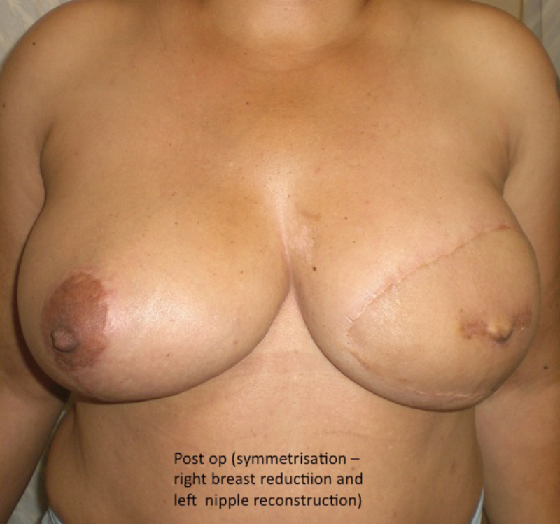
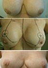
Figure 3 (above left): Post symmetrisation by right
breast reduction and left nipple reconstruction.
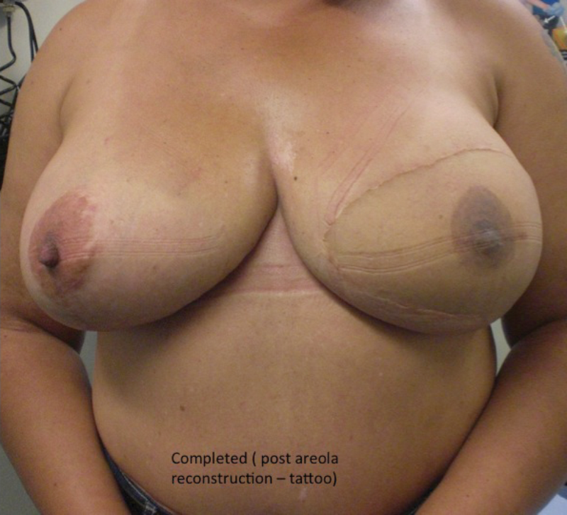
Figure 4 (above right): Completed – after reconstruction of the areola by tattoo.
Nipple reconstruction
The most common technique is by means of small flaps (star flaps, skate flaps, cervical visor (CV) flap, to mention a few commonly used techniques).
This is usually performed in conjunction with the symmetrisation. But I find it wise in a number of cases to perform this later as a short day case, when the symmetrisation procedure has settled and there is a satisfactory confirmation by both surgeon (me) and patient of acceptable symmetry outside a bra. This potentially may lead to a more symmetrical siting of the nipple areola complex. This is usually done without anaesthesia as the new breast mound is insensate but if need be a local anaesthetic will suffice.
It is important to emphasise to patients who have had implants to symmetrise the breasts, that the symmetry outside a bra is unlikely to be maintained for long as the breasts have different amounts of autologous and non-autologous tissue which cause different behaviour under gravity (droop at different rates) and time.
Stage 3: areola reconstruction
Areola reconstruction completes the process.
The most common method is by tattooing and this might require two or three episodes of tattooing to achieve a desired colour match with the opposite areola. I prefer this technique. Skin grafts, usually from the groin or the inner thighs, can also be used.
Timing of surgery (immediate versus delayed)
This is a decision between the oncological surgeon, plastic surgeon, oncologist, etc. and patient and tends to be ‘unit’ dependent. I use the word ‘unit’ to refer to the multidisciplinary and multiprofessional team that looks after the patient.
The debate about whether to perform immediate or delayed reconstruction continues, indicating that there is not a straightforward or correct answer. Most breast units have their policy or philosophy about the issue. Most are not evidence based and are a reflection of the idiosyncrasies of the clinicians and sometimes logistics of theatre space and operating times.
For patients who require significant adjuvant treatment, especially radiotherapy, most groups advise that the reconstruction is delayed as the radiotherapy can alter the result of the reconstruction. It must also be recognised that the priority is to treat the malignancy and any necessary adjuvant therapy should not be delayed in the event of surgical complications after the immediate reconstruction.
There are also a significant number of patients who just want to ‘get rid’ of the breast and decide later on if they want a reconstruction. Those who postpone their reconstruction go through a period of emotional re-adjustment to an absent breast mound and are likely to be more motivated if they seek reconstruction. There is also an argument that this subset of patients adjusts emotionally better after delayed reconstruction and ‘appreciates’ it more as they are comparing the reconstruction with no breast rather than with the lost breast. However, the cons include the paucity of the skin envelope and the possible fibrotic effects of post radiotherapy treatment on not only the skin but also the recipient blood vessels.
From my own experience and considering outcomes from a purely reconstructive perspective, immediate reconstructions are likely to give the best aesthetic results (especially in skin sparing mastectomy) as most of the breast skin is still available and not shrunken. In this case the reconstructive surgeon is basically ‘filling’ the cavity. The possible delay in inception of adjuvant treatment as a result of surgical complications and the negative effects of the adjuvant treatment, namely radiotherapy, are cons so an alternative strategy is to insert a temporary tissue expander at the time of mastectomy to minimise the skin shrinkage and maintain skin envelope for a later definitive reconstruction.
Recovery
Patients are discharged with a support bra to be worn day and night for six weeks except to have a shower or a bath. Patients who have had a DIEP flap are also fitted with an abdominal corset or binder for six weeks. These garments help support the tissue, reduce swelling and make the patient more comfortable.
Patients are seen weekly for wound check, review and physiotherapy. Patients are advised to avoid lifting (especially those who have young children) and to refrain from sexual activities for three to six weeks depending on the kind of reconstruction. For most patients, six weeks off work is recommended.
The reconstructed breast will not have normal sensation, although there may be restoration of some degree of protective feeling. It is important to warn patients of this in order to avoid accidental injury of the breast skin.
All scars are permanent. They can be improved with a good postoperative scar management.
Conclusion
Creating breasts that look natural in form, appearance and projection, and are symmetrical, at least within a bra, to the outside world should be the minimum of our objectives in breast reconstruction. There is no ‘ideal’ form of breast reconstruction for all patients and the choice of reconstruction depends on the individual circumstances. It is essential that the patient understands the process before it is begun.
There is usually a period of emotional re-adjustment to the new breast. Patients are generally very pleased they had the reconstruction. Their appearance and quality of life is improved. And reconstruction has not been shown to promote recurrence of breast cancer or prevent its detection.
In my opinion, breast reconstruction can be a most rewarding experience for both patient and surgeon. Every patient with a breast deformity should be aware of these advances in reconstructive surgery. For patients in the developing world there are distinct advantages in using their own (autologous) tissue and training surgeons in the developing world in these techniques should be encouraged and supported.
Declaration of competing interests: None declared.
COMMENTS ARE WELCOME